 Back
BackChapter 6: Bones and Bone Structure – Study Notes
Study Guide - Smart Notes
 Tailored notes based on your materials, expanded with key definitions, examples, and context.
Chapter 6: Bones and Bone Structure
Introduction to the Skeletal System
The skeletal system is a fundamental component of the human body, providing structure, support, and protection. It consists of bones, cartilages, ligaments, and other connective tissues that stabilize or interconnect the bones.
Structural Support: The skeleton forms the internal framework that supports the body and maintains its shape.
Mineral and Lipid Storage: Bones store essential minerals such as calcium (Ca) and phosphate, as well as lipids in yellow bone marrow.
Blood Cell Production: Red bone marrow produces red blood cells, white blood cells, and platelets.
Protection: Bones protect vital organs, including the brain, heart, and lungs.
Leverage for Movement: Bones act as levers that muscles pull on to produce movement.
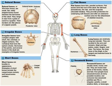
Classification of Bones by Shape
Bones are classified into six broad categories based on their shapes, each with distinct structural and functional characteristics.
Bone Type | Description | Examples |
|---|---|---|
Sutural (Wormian) Bones | Small, flat, irregularly shaped bones found between the flat bones of the skull. | Cranial sutures |
Irregular Bones | Complex shapes with short, flat, notched, or ridged surfaces. | Vertebrae, pelvic bones |
Short Bones | Boxlike in appearance, nearly equal in length and width. | Carpals (wrist), tarsals (ankle) |
Flat Bones | Thin with parallel surfaces, provide protection and surface area for muscle attachment. | Skull roof, sternum, ribs, scapulae |
Long Bones | Long and slender, found in limbs. | Humerus, femur, radius, ulna, tibia, fibula |
Sesamoid Bones | Small, round, and flat; develop inside tendons near joints. | Patella (kneecap), other variable locations |
Individual Variations
The number and location of sutural and sesamoid bones can vary among individuals, accounting for differences in total bone count.
The patella is the only named sesamoid bone; others are unnamed and variable.
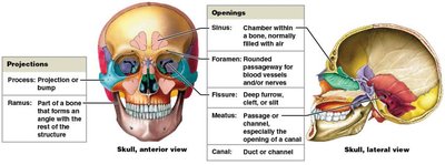
Bone Markings (Surface Features)
Bones have characteristic surface features called bone markings, which serve as sites for muscle, tendon, and ligament attachment, as well as passages for nerves and blood vessels.
Projections: Sites for muscle and ligament attachment or articulation with other bones (e.g., process, ramus, head, condyle, crest, tuberosity, trochanter).
Openings: Allow passage of blood vessels and nerves (e.g., foramen, canal, fissure, meatus, sinus).
Depressions: May accommodate soft tissues or form articulations (e.g., fossa, sulcus).
Type | Examples | Description |
|---|---|---|
Projection | Process, ramus, head, condyle, crest, tuberosity, trochanter | Attachment or articulation points |
Opening | Foramen, canal, fissure, meatus, sinus | Passageways for nerves/vessels |
Depression | Fossa, sulcus | Accommodate soft tissues or articulations |
Bone Structure
Structure of a Long Bone
Long bones, such as the femur, have a characteristic structure:
Diaphysis: The shaft, composed of compact bone surrounding a medullary (marrow) cavity.
Epiphysis: The expanded ends, consisting mainly of spongy (trabecular) bone covered by a thin layer of compact bone.
Metaphysis: The narrow region connecting the diaphysis to the epiphysis.
Structure of a Flat Bone
Flat bones, such as the parietal bone, consist of a layer of spongy bone (diploë) sandwiched between two layers of compact bone.
Bone Tissue and Matrix
Bone tissue is a specialized connective tissue with a dense matrix composed of calcium salts and collagen fibers.
Osteocytes: Mature bone cells located in lacunae.
Canaliculi: Narrow passageways connecting lacunae, allowing for nutrient and waste exchange.
Periosteum: A membrane covering the outer surface of bones, consisting of an outer fibrous layer and an inner cellular layer.
Bone Matrix Composition
About 2/3 of bone weight is calcium phosphate (), which interacts with calcium hydroxide () to form hydroxyapatite crystals ().
Hydroxyapatite incorporates other salts (e.g., calcium carbonate ) and ions (e.g., sodium, magnesium, fluoride).
About 1/3 of bone weight is collagen fibers, providing flexibility and a framework for mineral deposition.
The combination of collagen and hydroxyapatite gives bone both strength and some flexibility.
Bone Cells
There are four main types of bone cells, each with distinct functions:
Cell Type | Function | Location |
|---|---|---|
Osteogenic cells | Stem cells that differentiate into osteoblasts; important for fracture repair | Periosteum and endosteum |
Osteoblasts | Produce new bone matrix (osteoid) and promote mineralization | Bone surfaces |
Osteocytes | Mature bone cells; maintain bone matrix and help repair bone | Lacunae within bone matrix |
Osteoclasts | Resorb (break down) bone matrix; regulate calcium and phosphate levels | Bone surfaces (osteoclastic crypts) |
Osteoclasts are derived from the same stem cells as macrophages, not from osteogenic cells.
Compact and Spongy Bone
Compact Bone
The basic unit is the osteon (Haversian system), with a central canal containing blood vessels.
Lamellae: Layers of bone matrix; can be concentric (forming osteons), interstitial (between osteons), or circumferential (around the bone).
Perforating (Volkmann's) canals: Carry blood vessels perpendicular to the bone surface.
Spongy Bone
Composed of a network of trabeculae (no osteons).
Spaces between trabeculae contain red bone marrow (site of blood cell formation) or yellow bone marrow (fat storage).
Nutrients reach osteocytes by diffusion through canaliculi from blood vessels in the marrow.
Bone Membranes
Periosteum: Covers the outer surface of bones; isolates bone, provides a route for blood vessels and nerves, and participates in growth and repair.
Endosteum: Lines the inner surfaces of bone (medullary cavity, trabeculae, central canals); active in bone growth, repair, and remodeling.
Bone Formation and Growth
Ossification (Osteogenesis)
Ossification is the process of bone formation. There are two main types:
Endochondral Ossification: Bone replaces a pre-existing cartilage model; most bones, especially long bones, form this way.
Intramembranous Ossification: Bone develops directly from mesenchymal tissue; forms most flat bones.
Endochondral Ossification Steps
Chondrocytes enlarge and the cartilage matrix calcifies, causing cell death and cavity formation.
Blood vessels invade the perichondrium, converting it to periosteum; osteoblasts form a bone collar.
Blood vessels penetrate the cartilage, bringing osteoblasts that form spongy bone at the primary ossification center.
Remodeling creates a medullary cavity; bone formation spreads toward the epiphyses.
Secondary ossification centers form in the epiphyses.
Epiphyses fill with spongy bone; the epiphyseal plate (cartilage) separates epiphysis from diaphysis.
At puberty, ossification outpaces cartilage growth, leading to epiphyseal closure and formation of the epiphyseal line.
Appositional Growth: Bones grow in width by adding new bone at the surface.
Intramembranous Ossification Steps
Mesenchymal cells differentiate into osteoblasts, which secrete osteoid that calcifies to form bone matrix.
Osteoblasts become osteocytes as they are surrounded by matrix; bone grows in spicules.
Blood vessels grow into the area, accelerating bone growth.
Spongy bone forms; outer layers are remodeled into compact bone, and periosteum develops.
Bone Vascularization and Innervation
Bones are highly vascular, with nutrient arteries and veins supplying the diaphysis, metaphyseal vessels supplying the metaphysis, and periosteal vessels supplying the outer layers.
The periosteum contains lymphatic vessels and sensory nerves, making bone injuries painful.
Bone Remodeling
Bone remodeling is a continuous process involving osteocytes, osteoblasts, and osteoclasts. It allows bones to adapt to stress and repair microdamage.
Osteoclasts resorb bone matrix; osteoblasts deposit new bone.
Balance between these activities determines bone strength.
Physical activity increases bone strength; inactivity or excessive osteoclast activity weakens bones.
Calcium Homeostasis
The skeleton acts as a reservoir for calcium, which is vital for nerve and muscle function. Blood calcium levels are tightly regulated by hormones.
Parathyroid Hormone (PTH): Released when blood calcium is low; increases osteoclast activity, enhances intestinal absorption (via calcitriol), and decreases renal excretion of calcium.
Calcitonin: Released when blood calcium is high; inhibits osteoclasts, increases renal excretion, and decreases intestinal absorption of calcium.
Fractures
A fracture is a break or crack in a bone caused by physical stress.
Open (Compound) Fracture: Bone projects through the skin.
Closed (Simple) Fracture: Bone does not penetrate the skin.
