 Back
BackChapter 6: Bones and Skeletal Tissues – Comprehensive Study Notes
Study Guide - Smart Notes
 Tailored notes based on your materials, expanded with key definitions, examples, and context.
Bones and Skeletal Tissues
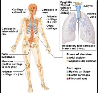
Cartilage: Basic Structure, Types, and Locations
Cartilage is a resilient, flexible connective tissue found throughout the body, especially in areas requiring cushioning and support. It is avascular and lacks nerves, making it distinct from bone.
Skeletal cartilage: Contains chondrocytes in lacunae and an extracellular matrix. Surrounded by the perichondrium, which provides nutrients and resists expansion.
Types of cartilage:
Hyaline cartilage: Most abundant; provides support, flexibility, and resilience. Found in articular, costal, respiratory, and nasal cartilages.
Elastic cartilage: Similar to hyaline but contains elastic fibers; found in the external ear and epiglottis.
Fibrocartilage: Contains thick collagen fibers; provides tensile strength. Located in menisci of the knee and intervertebral discs.
Growth types:
Appositional growth: Cells secrete matrix on the external face of cartilage.
Interstitial growth: Chondroblasts secrete new matrix from within.
Calcification: Occurs during bone growth but calcified cartilage is not bone.
Comparison of Cartilage and Bone
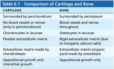
Cartilage and bone are both connective tissues but differ in structure, function, and growth.
Cartilage | Bone |
|---|---|
Surrounded by perichondrium | Surrounded by periosteum |
No blood vessels or nerves (except in perichondrium) | Blood vessels and nerves throughout |
Chondrocytes in lacunae | Osteocytes in lacunae |
Flexible extracellular matrix | Rigid extracellular matrix (due to inorganic calcium salts) |
Matrix made by chondroblasts | Matrix (organic part) made by osteoblasts |
Appositional and interstitial growth | Appositional growth only |
Classification of Bones by Location and Shape
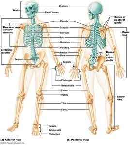
The human skeleton consists of 206 named bones, classified by their location and shape.
Axial skeleton: Long axis of the body; includes skull, vertebral column, and rib cage.
Appendicular skeleton: Bones of limbs and girdles attaching limbs to the axial skeleton.
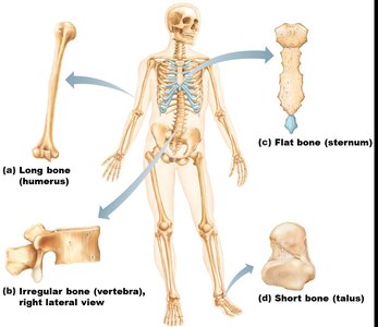
Bone shapes:
Long bones: Longer than wide (e.g., humerus).
Short bones: Cube-shaped (e.g., wrist, ankle); sesamoid bones (e.g., patella).
Flat bones: Thin, flat, slightly curved (e.g., sternum, ribs, skull).
Irregular bones: Complicated shapes (e.g., vertebrae, coxal bones).
Functions of the Skeletal System
The skeletal system performs several essential functions for the human body:
Support: Provides structural framework for the body and soft organs.
Protection: Shields the brain, spinal cord, and vital organs.
Movement: Acts as levers for muscle action.
Mineral storage: Reservoir for calcium, phosphorus, and growth factors.
Blood cell formation: Hematopoiesis occurs in red marrow cavities.
Triglyceride storage: Fat stored in bone cavities as an energy source.
Hormone production: Osteocalcin regulates insulin secretion and protects against obesity and diabetes.
Structure of Bones
Bones are organs composed of various tissues, including bone (osseous) tissue, nervous tissue, cartilage, fibrous connective tissue, muscle, and epithelial cells in blood vessels. Bone structure is studied at three levels: gross anatomy, microscopic, and chemical.
Bone Textures: Compact and Spongy Bone
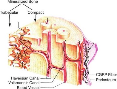
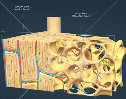
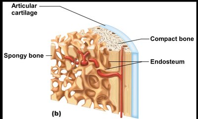
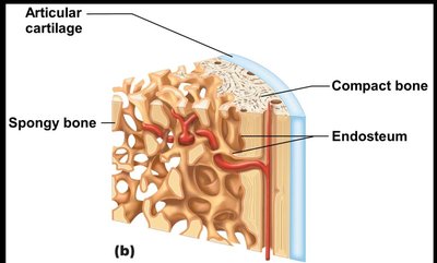
Compact bone: Dense outer layer; smooth and solid.
Spongy bone (cancellous or trabecular): Honeycomb of flat pieces called trabeculae, found deep to compact bone.
Gross Anatomy: Structure of Long Bones
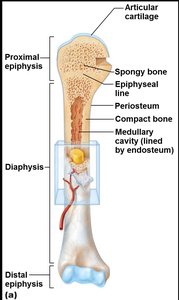
Diaphysis: Tubular shaft forming the long axis; compact bone surrounds the medullary cavity.
Epiphyses: Bone ends; external compact bone, internal spongy bone, articular cartilage covers surfaces.
Epiphyseal line: Remnant of childhood bone growth at the epiphyseal plate.
Gross Anatomy: Membranes
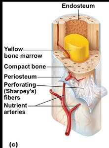
Periosteum: White, double-layered membrane covering external surfaces except joints; contains nerve fibers, blood vessels, and osteogenic cells.
Endosteum: Delicate membrane covering internal bone surfaces, trabeculae of spongy bone, and lining canals in compact bone; contains osteogenic cells.
Bone Cavities: Red and Yellow Marrow
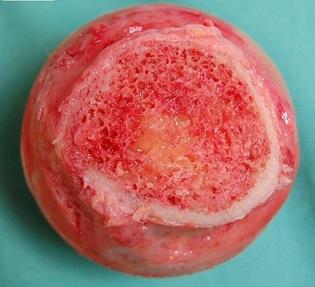
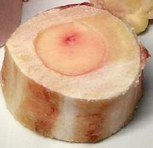
Red marrow: Found in trabecular cavities of spongy bone in flat bones and heads of long bones; site of hematopoiesis.
Yellow marrow: Fat storage in medullary cavity; can convert to red marrow if necessary.
Bone Markings
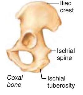
Projections: Sites for muscle and ligament attachment (e.g., iliac crest, ischial tuberosity).
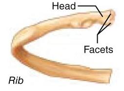
Surfaces: Areas forming joints (e.g., head, facets).
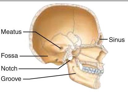
Depressions and openings: Conduits for blood vessels and nerves (e.g., meatus, fossa, notch, groove, sinus, foramen).
Microscopic Anatomy of Bone: Cells of Bone Tissue
Bone tissue contains five major cell types, each with specialized functions:
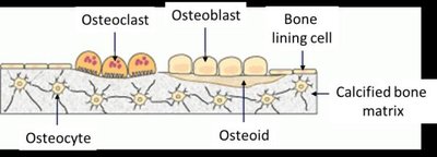
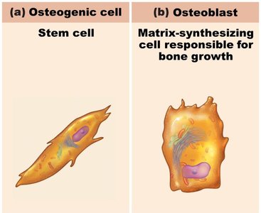
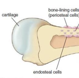
Osteogenic cells: Stem cells in periosteum and endosteum; differentiate into osteoblasts or bone lining cells.
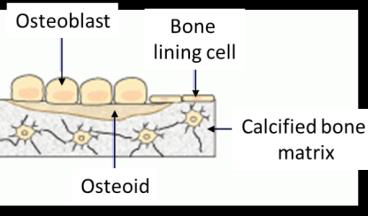
Osteoblasts: Bone-forming cells; secrete unmineralized bone matrix (osteoid) including collagen and calcium-binding proteins.
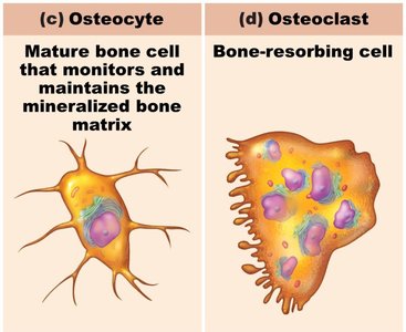
Osteocytes: Mature bone cells in lacunae; monitor and maintain bone matrix, act as stress sensors.
Bone lining cells: Flat cells on bone surfaces; help maintain matrix (periosteal and endosteal cells).
Osteoclasts: Derived from hematopoietic stem cells; giant, multinucleate cells for bone resorption.
Microscopic Anatomy: Compact Bone
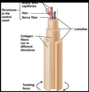
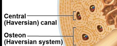
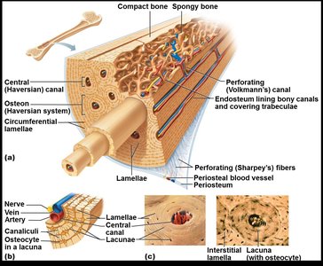
Osteon (Haversian system): Structural unit; elongated cylinder parallel to bone axis, composed of concentric lamellae.
Lamellae: Collagen fibers in adjacent rings run in different directions, resisting twisting forces.
Canals and canaliculi:
Central (Haversian) canal: Contains blood vessels and nerve fibers.
Perforating (Volkmann's) canals: Connect blood vessels and nerves of periosteum, medullary cavity, and central canal.
Canaliculi: Hairlike canals connecting lacunae; allow communication and nutrient/waste exchange between osteocytes.
Microscopic Anatomy: Spongy Bone
Trabeculae: Align along lines of stress; contain irregularly arranged lamellae and osteocytes interconnected by canaliculi.
No osteons; capillaries in endosteum supply nutrients.
Chemical Composition of Bone
Organic components: Cells and osteoid (ground substance and collagen fibers); provide tensile strength and flexibility.
Inorganic components: Hydroxyapatites (mineral salts, mainly calcium phosphate); responsible for hardness and resistance to compression.
Bone Development (Ossification)
Ossification is the process of bone tissue formation, beginning in the second month of development and continuing throughout life.
Formation of bony skeleton:
Endochondral ossification: Bone forms by replacing hyaline cartilage; forms most of the skeleton.
Intramembranous ossification: Bone develops from fibrous membrane; forms flat bones (clavicles, cranial bones).
Postnatal bone growth:
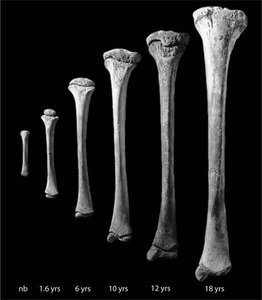
Interstitial growth: Increase in length of long bones.
Appositional growth: Increase in bone thickness.
Bone remodeling and repair: Lifelong process involving appositional growth.
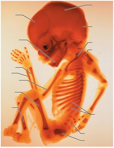
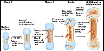
Steps in Endochondral Ossification
Primary ossification center forms in the shaft.
Blood vessel infiltration converts perichondrium to periosteum; cells become osteoblasts.
Bone collar forms around diaphysis of cartilage model.
Central cartilage in diaphysis calcifies and develops cavities.
Periosteal bud invades cavities, forming spongy bone.
Diaphysis elongates and medullary cavity forms.
Epiphyses ossify.
Steps in Intramembranous Ossification
Mesenchymal cells cluster and differentiate into osteoblasts, forming ossification centers.
Osteoblasts secrete osteoid, which calcifies; osteoblasts become osteocytes within lacunae.
Osteoid is laid down around blood vessels, forming immature trabeculae; mesenchyme condenses to form periosteum.
Trabeculae below periosteum are remodeled into compact bone; space is filled with red marrow.
Postnatal Bone Growth
Interstitial (longitudinal) growth: Requires epiphyseal cartilage; epiphyseal plate maintains constant thickness.
Appositional growth: Osteoblasts secrete matrix on external bone; osteoclasts remove bone on endosteal surface.
Epiphyseal Plate Zones
Resting (quiescent) zone: Relatively inactive cartilage.
Proliferation (growth) zone: Rapidly dividing cartilage cells lengthen bone.
Hypertrophic zone: Older chondrocytes enlarge and erode, creating spaces.
Calcification zone: Matrix calcifies, chondrocytes die.
Ossification zone: Calcified cartilage replaced by spongy bone.
Epiphyseal Plate Closure
Near end of adolescence, chondroblasts divide less often; plate thins and is replaced by bone (epiphyseal line).
Bone lengthening ceases; epiphysis and diaphysis fuse (females ~18 years, males ~21 years).
Appositional Growth: Growth in Width
Allows bones to widen throughout life.
Osteoblasts build bone externally; osteoclasts remove bone internally.
Results in thicker, stronger bones.
Bone Homeostasis
Bone homeostasis involves the continuous recycling of bone mass through remodeling and repair.
Spongy bone replaced every 3-4 years; compact bone every 10 years.
Older bone becomes brittle due to crystallization of calcium salts.
Remodeling occurs at periosteum and endosteum surfaces; osteoblasts build bone, osteoclasts break down bone.
Mechanical stress determines where remodeling occurs; hormonal controls determine when.
Control of Remodeling and Importance of Calcium
Remodeling regulated by genetic factors and two control pathways:
Negative feedback hormonal loop for Ca2+ homeostasis (parathyroid hormone, PTH).
Responses to mechanical and gravitational forces.
Calcium is essential for nerve and muscle transmission, bone strength, blood coagulation, secretion, and cell division.
99% of body calcium is stored in bones; blood levels tightly regulated (9-11 mg/dl).
Negative Feedback Hormonal Loop for Blood Ca2+ Homeostasis
Low blood Ca2+ triggers PTH release.
PTH stimulates osteoclasts to degrade bone matrix, releasing Ca2+.
Blood Ca2+ levels rise; PTH release ends.
Bone Repair
Fractures are breaks in bone; most result from trauma in youth or bone thinning in old age.
Treatment involves reduction (realignment), immobilization, and healing.
Repair and healing occur in four steps.
Bone Disorders
Osteoporosis: Bone-thinning disease; common in older adults, especially women. Bones become fragile and fracture easily.
Osteomalacia: Poorly mineralized (soft) bones due to insufficient calcium or vitamin D; called Rickets in children, causing deformities.
Paget's Disease: Haphazard bone deposition and resorption; reduced mineralization, excessive spongy bone, spotty weakening.
Bony spurs: Abnormal bone projections due to overgrowth; common in aging bones.
Osteosarcoma: Bone cancer (osteoblasts); aggressive, most common in young adults, often metastasizes to lungs.
Summary of Learning Objectives
Identify and describe characteristics of skeletal cartilage, types of growth, and differences from bone.
Classify bones by location and shape.
List the seven functions of the skeletal system.
Understand why bones are tissues and list three levels of bone structure.
Describe gross anatomical characteristics of bones: textures, structures, membranes, cavities, and markings.
Describe microscopic characteristics: cells and features of compact and spongy bone.
Understand chemical composition and features provided to bones.
Know processes of ossification, postnatal growth, and bone remodeling/repair.
Describe bone homeostasis, remodeling causes, and calcium's role.
List types of bone fractures and steps in bone repair.
Describe characteristics of five bone disorders.
