 Back
BackChapter 6: Bones and Skeletal Tissues – Structured Study Notes
Study Guide - Smart Notes
 Tailored notes based on your materials, expanded with key definitions, examples, and context.
Chapter 6: Bones and Skeletal Tissues
Skeletal Cartilage and Bone Foundations
The skeletal system is built upon a foundation of cartilage, which later becomes bone through ossification. Cartilage is avascular and lacks nerves, making it distinct from bone tissue.
Types of Cartilage: Hyaline (most common, provides support and flexibility), Elastic (more flexible, found in ear and epiglottis), Fibrous (high tensile strength, found in intervertebral discs).
Perichondrium: Dense connective tissue surrounding cartilage; provides nutrients and support.
Cartilage Growth:
Appositional: Growth in length; new cells added to the surface.
Interstitial: Growth from within; cells divide and expand the tissue internally.
Chondroblasts: Immature cells responsible for cartilage growth.
Chondrocytes: Mature cells that maintain cartilage structure.
Functions of Bone
Bones serve multiple essential functions in the human body, contributing to both structure and physiology.
Support: Framework for the body.
Protection: Shields vital organs (e.g., skull protects brain, rib cage protects heart and lungs).
Movement: Muscles attach to bones, enabling movement.
Mineral Storage: Stores calcium, magnesium, potassium.
Blood Cell Formation: Hematopoiesis occurs in bone marrow.
Fat Storage: Yellow marrow stores fat.
Hormone Production: Bones produce hormones such as osteocalcin.
Classification of Bones
Bones are classified by their location and shape, which reflects their function and structure.
Total Bones: 206 in the adult human body, mostly paired.
Axial Skeleton: Head, vertebral column, rib cage.
Appendicular Skeleton: Limbs (upper and lower).
Shapes:
Long Bones: Rectangular shape (e.g., humerus, finger bones).
Short Bones: Cube-like (e.g., carpals).
Flat Bones: Thin, flat (e.g., sternum).
Irregular Bones: Complex shapes (e.g., pelvis, vertebrae).
Bone Structure and Gross Anatomy
Bones are composed of several tissue types and have distinct anatomical regions.
Osseous Tissue: Main bone tissue.
Nerves, Cartilage, Fibrous Connective Tissue, Epithelial Cells: Contribute to bone function and structure.
Compact Bone: Dense outer layer.
Spongy Bone: Porous inner layer.
Periosteum: Outer covering of bone.
Endosteum: Lines internal surfaces.
Long Bone Anatomy
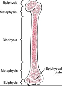
Long bones have a distinct structure, including diaphysis, epiphyses, and metaphyses.
Diaphysis: Shaft; contains medullary cavity (marrow, fat).
Epiphyses: Ends; more spongy bone, less compact bone.
Epiphyseal Plate/Line: Site of lengthwise growth (appositional growth).
Hematopoietic Tissue
Bone marrow is the site of blood cell formation (hematopoiesis).
Red Marrow: Found in spongy bone of flat bones; in infants, present in all bones; in adults, limited to heads of long bones.
Bone Markings
Bones have markings that serve as sites for muscle, ligament, and tendon attachment, as well as passageways for nerves and blood vessels.
Projections: Bulges or protrusions for attachment.
Depressions: Indentations that house nerves and blood vessels.
Openings: Holes or canals for passage of nerves and blood vessels.
Microscopic Anatomy of Bone
Bone tissue contains several specialized cell types, each with distinct functions.
Osteogenic Cells: Stem cells; differentiate into osteoblasts.
Osteoblasts: Bone-forming cells; secrete matrix (osteoid).
Osteocytes: Mature bone cells; maintain matrix; reside in lacunae.
Bone Lining Cells: Maintain matrix; periosteal and endosteal cells.
Osteoclasts: Bone-resorbing cells; break down bone tissue.
Compact Bone Structure
Osteon: Structural unit; concentric lamellae.
Central Canal: Houses blood vessels and nerves.
Lacunae: Spaces housing osteocytes.
Canaliculi: Channels for communication between lacunae.
Interstitial Lamellae: Between osteons.
Circumferential Lamellae: Between endosteum and periosteum.
Spongy Bone Structure
Trabeculae: Porous network; no osteons.
Ossification (Bone Formation)
Ossification is the process of bone formation, beginning in the embryo and continuing into early adulthood.
Endochondral Ossification: Replaces hyaline cartilage; forms most bones below the skull (except clavicle).
Intramembranous Ossification: Replaces fibrous tissue; forms skull bones and clavicle.
Endochondral Ossification Steps
Bone collar forms around diaphysis.
Cartilage calcifies and forms cavities.
Periosteal bud invades, forming spongy bone.
Diaphysis elongates; secondary ossification centers form in epiphyses.
Intramembranous Ossification Steps
Ossification centers form in fibrous tissue.
Osteoblasts secrete osteoid, which calcifies.
Trabeculae form around blood vessels.
Spongy bone develops with red bone marrow.
Bone Growth
Bones grow in length and width through distinct processes regulated by hormones and physical activity.
Interstitial Growth: Lengthwise growth at epiphyseal plate; involves five zones: resting, proliferation, hypertrophic, calcification, ossification.
Appositional Growth: Widthwise growth throughout life; stimulated by physical stress.
Hormonal Regulation: Growth hormone, thyroid hormone, testosterone, parathyroid hormone, and calcitonin all influence bone growth and remodeling.
Bone Remodeling
Bone remodeling is a lifelong process involving deposition and resorption, regulated by osteoblasts and osteoclasts.
Deposition: Osteoblasts add new bone tissue.
Resorption: Osteoclasts break down bone tissue.
Wolff’s Law: Bone adapts to physical stresses and hormonal demands.
Fractures and Repair
Bone fractures are classified by position, completeness, and skin involvement. Repair involves several stages.
Types: Displaced vs. nondisplaced; complete vs. incomplete; open vs. closed.
Repair Methods: Closed reduction (manual), open reduction (surgical), casting/immobilization.
Stages of Repair:
Hematoma formation (blood clot).
Fibrocartilage callus formation (capillaries return, debris removed, collagen fibers connect ends).
Bony callus formation (new trabeculae, compact bone returns).
Bone remodeling.
Bone Disorders
Several disorders affect bone health and structure.
Osteomalacia: Softening of bone due to decreased minerals.
Rickets: Softening in children due to low vitamin D or calcium; causes bowed legs.
Osteoporosis: Decreased bone density; common in menopausal women due to lack of estrogen. Resorption exceeds deposition.
Paget’s Disease: Excessive bone growth; most common in adults over 40; etiology unknown.
Additional info: Hormonal regulation and physical activity are critical for maintaining bone health. Estrogen supplements, weight-bearing exercise, and adequate calcium/vitamin D intake are recommended for prevention and management of osteoporosis.
