 Back
BackChapter 6: Communication, Integration, and Homeostasis – Study Notes
Study Guide - Smart Notes
 Tailored notes based on your materials, expanded with key definitions, examples, and context.
Communication, Integration, and Homeostasis
Introduction
This chapter explores the mechanisms by which cells communicate, integrate signals, and maintain homeostasis in the human body. Understanding these processes is fundamental to physiology, as they underlie the regulation of all organ systems.
Cell-to-Cell Communication
Types of Physiological Signals
Electrical signals: Changes in the membrane potential of a cell, crucial for nerve and muscle function.
Chemical signals: Molecules such as hormones, neurotransmitters, and cytokines secreted into the extracellular fluid (ECF) to mediate most communication within the body. These signals are called ligands.
Protein binding rules: Ligand-receptor interactions exhibit specificity, affinity, competition, and saturation.
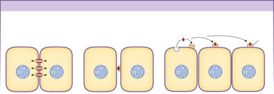
Local Communication
Gap junctions: Direct cytoplasmic connections between adjacent cells, allowing ions and small molecules to pass freely.
Contact-dependent signals: Require direct interaction between membrane molecules on two cells.
Autocrine signals: Act on the same cell that secreted them.
Paracrine signals: Secreted by one cell and diffuse to adjacent cells.
Long-Distance Communication
Hormones: Secreted by endocrine cells into the blood; only target cells with specific receptors respond.
Neurotransmitters: Chemicals secreted by neurons that diffuse across a small gap to the target cell.
Neurohormones: Chemicals released by neurons into the blood for action at distant targets.
Cytokines
Cytokines: Peptides synthesized and secreted by all nucleated cells in response to stimuli. They can act as autocrine, paracrine, or long-distance signals, especially in immune responses and development.
Signal Pathways
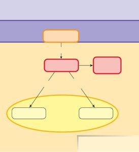
General Steps in Signal Transduction
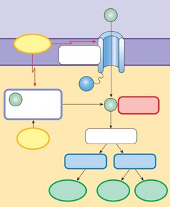
The chemical signal (first messenger) binds to a receptor protein.
Ligand-receptor binding activates the receptor.
The receptor activates one or more intracellular signal molecules (second messengers).
The last signal molecule modifies existing proteins or initiates the synthesis of new proteins.
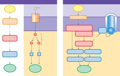
Receptor Locations
Intracellular receptors: For lipophilic signals (e.g., steroid hormones) that diffuse through the cell membrane and bind to cytosolic or nuclear receptors, often affecting gene expression (slower response).
Cell membrane receptors: For lipophobic signals (e.g., peptide hormones) that cannot cross the membrane and bind to receptors on the cell surface (rapid response).
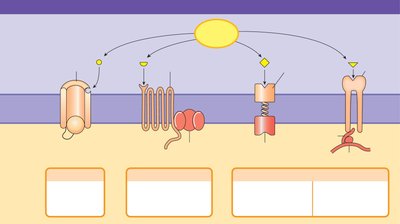
Categories of Membrane Receptors
Chemically gated (ligand-gated) ion channels: Open or close in response to ligand binding.
G protein-coupled receptors (GPCRs): Activate intracellular signaling cascades via G proteins.
Receptor-enzymes: Have intrinsic enzyme activity or are associated with enzymes.
Integrin receptors: Involved in cell adhesion and signaling to the cytoskeleton.
Signal Transduction Mechanisms
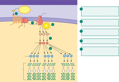
Signal transduction: The process by which an extracellular signal is converted into an intracellular response, often involving protein kinases and amplifier enzymes.
First messenger: The extracellular signal molecule.
Second messenger: Intracellular molecules that propagate the signal (e.g., cAMP, Ca2+).
Amplification: A single signal molecule can generate a large intracellular response via amplifier enzymes.
G Protein-Coupled Receptor (GPCR) Pathways
GPCRs activate G proteins, which can turn on amplifier enzymes such as adenylyl cyclase.
Adenylyl cyclase converts ATP to cAMP, which activates protein kinase A, leading to phosphorylation of proteins and a cellular response.
Second Messenger Pathways
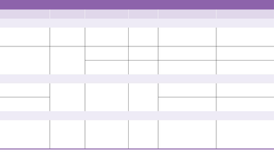
Second Messenger | Made from | Amplifier Enzyme | Linked to | Action/Effects |
|---|---|---|---|---|
cAMP | ATP | Adenylyl cyclase | GPCR | Activates protein kinases (PKA), binds ion channels, alters channel opening |
cGMP | GTP | Guanylyl cyclase | Receptor-enzyme | Activates protein kinases (PKG), phosphorylates proteins |
IP3 | Membrane phospholipids | Phospholipase C | GPCR | Releases Ca2+ from intracellular stores |
DAG | Membrane phospholipids | Phospholipase C | GPCR | Activates protein kinase C, phosphorylates proteins |
Ca2+ | Extracellular fluid or intracellular stores | — | — | Binds to calmodulin and other proteins, alters enzyme activity, exocytosis, muscle contraction |
Rapid Signal Pathways
Ligand-gated ion channels are found mostly in nerve and muscle tissue.
Ligand binding changes ion permeability, causing rapid cellular responses such as depolarization.
Novel Signal Molecules
Calcium as a Second Messenger
Calcium enters cells through voltage, ligand, or mechanically gated channels.
It binds to calmodulin or other regulatory proteins, altering protein activity, triggering exocytosis, or initiating movement.
Gaseous Signal Molecules
Nitric oxide (NO): Produced by endothelial cells, diffuses into smooth muscle, and causes vasodilation. Acts as a short-acting paracrine or autocrine signal.
Lipid-Derived Paracrine Signals
Arachidonic acid cascade: Membrane phospholipids are converted to arachidonic acid, which is further metabolized to eicosanoids (e.g., prostaglandins, leukotrienes, thromboxanes) involved in inflammation, pain, blood clotting, and fever.
Modulation of Signal Pathways
Receptor Properties
Specificity: Receptors bind only specific ligands.
Saturation: Maximum response occurs when all receptors are occupied.
Competition: Multiple ligands may compete for the same receptor (e.g., norepinephrine and epinephrine for adrenergic receptors).
Agonists: Ligands that activate receptors and elicit a response.
Antagonists: Ligands that bind receptors but block activation by the primary ligand.
Receptor Isoforms and Target Response
One ligand may have multiple receptor isoforms, leading to different cellular responses depending on the receptor type.
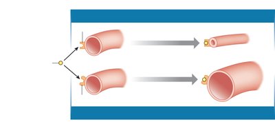
Example: Epinephrine causes vasoconstriction in intestinal blood vessels (α-receptor) and vasodilation in skeletal muscle blood vessels (β2-receptor).
Regulation of Receptor Number
Down-regulation: Decrease in receptor number, reducing cell sensitivity to a signal.
Up-regulation: Increase in receptor number, enhancing cell sensitivity.
Many drugs target signal transduction proteins (e.g., beta blockers for blood pressure control).
Homeostatic Reflex Pathways
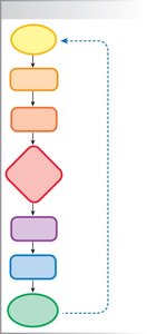
Components of a Reflex Pathway
Input: Stimulus is detected by a sensor or receptor, which generates an input signal (afferent pathway).
Integration: Integrating center compares the input with a setpoint.
Output: Output signal (efferent pathway) is sent to the target (effector organ), producing a response.
Nervous vs. Endocrine Control Systems
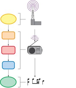
Speed: Neural control is faster than endocrine control.
Duration: Neural responses are shorter in duration; endocrine responses last longer.
Coding for stimulus intensity: Neural systems use frequency of action potentials; endocrine systems use the amount of hormone released.
Summary Table: Key Concepts in Communication, Integration, and Homeostasis
Topic | Main Points |
|---|---|
Cell-to-Cell Communication | Electrical and chemical signals, local and long-distance communication, cytokines |
Signal Pathways | Receptors, signal transduction, second messengers, amplification |
Novel Signal Molecules | Calcium, gases (NO), lipid-derived signals (eicosanoids) |
Modulation of Pathways | Specificity, competition, agonists/antagonists, receptor regulation |
Homeostatic Reflexes | Input, integration, output, nervous vs. endocrine control |
