 Back
Chapter 6: The Skeletal System – Structure, Function, and Organization
Study Guide - Smart Notes
 Tailored notes based on your materials, expanded with key definitions, examples, and context.
The Skeletal System
Overview and Components
The skeletal system is a complex framework that provides support, protection, and movement for the human body. It consists of bones, cartilages, joints, ligaments, and connective tissues that stabilize or connect bones.
Bones: Rigid organs that form the skeleton.
Cartilages: Flexible connective tissues at joints.
Joints: Articulations where bones meet.
Ligaments: Connect bones to other bones.
Connective tissues: Stabilize and support skeletal elements.
Primary Functions of the Skeletal System
Support: Provides structural support for the entire body and attachment points for soft tissues and organs.
Storage of Minerals and Lipids: Stores calcium and phosphorus ions in bone matrix; yellow bone marrow stores lipids for energy.
Blood Cell Production: Red bone marrow produces red blood cells, white blood cells, and platelets.
Protection: Surrounds and protects vital organs (e.g., skull protects brain, ribs protect heart and lungs).
Leverage: Bones act as levers to change the direction and magnitude of muscle forces, enabling movement.
Bone Tissue and Structure
Bone (Osseous) Tissue
Bone tissue is a specialized connective tissue composed of cells (osteocytes) embedded in a mineralized matrix.
Matrix: Contains calcium phosphate (Ca3(PO4)2) and collagen fibers.
Osteocytes: Mature bone cells located in lacunae, maintaining bone matrix.
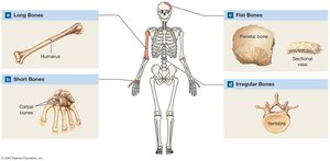
Classification of Bones by Shape
Bones are classified based on their shapes, which relate to their functions.
Long bones: Longer than wide (e.g., humerus).
Short bones: About as wide as long (e.g., carpal bones).
Flat bones: Thin and broad (e.g., parietal bone).
Irregular bones: Complex shapes (e.g., vertebrae).
Features of a Long Bone
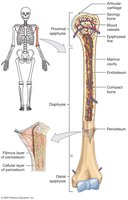
Diaphysis: Central shaft surrounding the marrow (medullary) cavity.
Epiphyses: Expanded ends covered with articular cartilage, forming joints.
Periosteum: Outer covering with fibrous and cellular layers; isolates bone and provides attachment for tendons/ligaments.
Endosteum: Lines marrow cavity and spongy bone; active in bone growth and repair.
Types of Bone Tissue
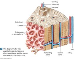
Compact bone (dense bone): Forms the wall of the diaphysis; solid and strong.
Spongy bone (cancellous bone): Network of trabeculae; found in epiphyses and lines marrow cavity; lighter and reduces bone weight.
Microscopic Structure of Bone
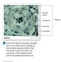
Osteon (Haversian system): Basic unit of compact bone; concentric lamellae around a central canal containing blood vessels.
Lacunae: Small spaces housing osteocytes.
Canaliculi: Tiny channels connecting lacunae for nutrient/waste exchange.
Types of Bone Cells
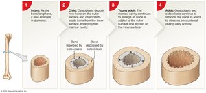
Osteoblasts: Bone-building cells; synthesize new bone matrix (ossification).
Osteocytes: Mature bone cells; maintain bone matrix.
Osteoclasts: Bone-resorbing cells; break down bone matrix, releasing minerals (resorption).
Bone Formation and Growth
Ossification Processes
Bone formation (ossification) replaces other tissues with bone during development. Two main processes:
Intramembranous ossification: Bone develops from fibrous connective tissue (e.g., flat bones of skull).
Endochondral ossification: Bone replaces hyaline cartilage models (most bones).
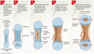
Appositional Growth
Bone increases in diameter through appositional growth, where new bone is added to the outer surface by osteoblasts, and osteoclasts enlarge the marrow cavity from within.
Requirements for Bone Growth
Minerals: Calcium and phosphate are essential for bone matrix.
Vitamin D3: Needed for calcium absorption; deficiency leads to osteomalacia or rickets.
Vitamins A and C: Support osteoblast activity.
Hormones: Growth hormone, thyroid hormone, sex hormones, parathyroid hormone (PTH), and calcitonin regulate bone growth and calcium balance.
Bone Remodeling and Repair
Bone Remodeling
Bone remodeling is a continuous process where old bone is replaced by new bone, allowing adaptation to stress and repair of microdamage.
Osteocytes maintain matrix; osteoclasts resorb bone; osteoblasts form new bone.
Exercise increases bone strength; inactivity leads to bone loss.
Calcium Homeostasis
Bone acts as a calcium reserve, with 99% of body calcium stored in the skeleton. Blood calcium levels are tightly regulated by:
Parathyroid hormone (PTH) and calcitriol: Increase blood calcium by stimulating bone resorption.
Calcitonin: Lowers blood calcium by inhibiting bone resorption.
Fracture Repair
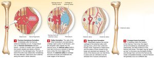
Bone fractures are classified as closed (simple) or open (compound). Repair involves:
Formation of a fracture hematoma (blood clot).
Callus formation by periosteum and endosteum cells.
Replacement of cartilage with spongy bone by osteoblasts.
Remodeling of spongy bone into compact bone.
Organization of the Skeleton
Axial and Appendicular Skeletons
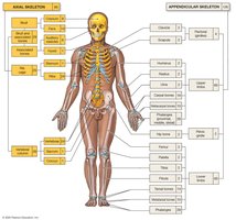
The human skeleton is divided into two main parts:
Axial skeleton: Skull, vertebral column, and thoracic cage (80 bones).
Appendicular skeleton: Limbs and girdles (126 bones).
Bone Markings
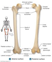
Bone markings are surface features that serve as attachment points for muscles, tendons, and ligaments, or as passages for nerves and blood vessels.
Elevations/Projections: Sites of attachment or articulation.
Depressions/Openings: Passageways for nerves and vessels.
Axial Skeleton: Skull, Vertebral Column, and Thoracic Cage
Skull
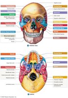
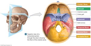
Cranial bones: Protect the brain (e.g., frontal, parietal, occipital, temporal, sphenoid, ethmoid).
Facial bones: Form the face and support entrances to digestive and respiratory tracts (e.g., maxilla, mandible, zygomatic, nasal).
Paranasal Sinuses and Nasal Complex
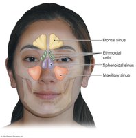
Paranasal sinuses are air-filled spaces that lighten the skull and produce mucus. The nasal complex includes bones and cartilage forming the nasal cavity and septum.
Hyoid Bone and Infant Skull
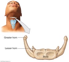
Hyoid bone: U-shaped bone in the neck; supports the larynx and tongue.
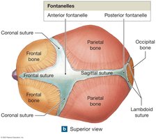
Fontanelles: Soft spots in infant skulls; allow for brain growth and easier birth.
Vertebral Column
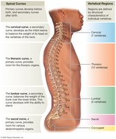
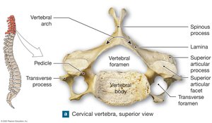
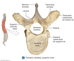
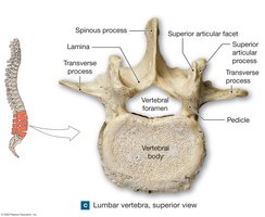
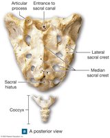
The vertebral column consists of 26 bones: 24 vertebrae, the sacrum, and the coccyx. It supports the body and protects the spinal cord.
Cervical (7), Thoracic (12), Lumbar (5), Sacrum (5 fused), Coccyx (3-5 fused)
Thoracic Cage
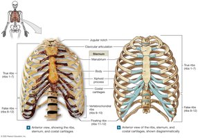
The thoracic cage consists of the thoracic vertebrae, ribs, and sternum. It protects the heart and lungs and supports the shoulder girdles.
True ribs (1-7): Attach directly to sternum.
False ribs (8-12): Attach indirectly or not at all.
Floating ribs (11-12): No anterior attachment.
Appendicular Skeleton: Limbs and Girdles
Pectoral Girdle and Upper Limb
Pectoral girdle: Clavicle and scapula; connects upper limbs to trunk.
Upper limb: Humerus (arm), radius and ulna (forearm), carpals (wrist), metacarpals (hand), phalanges (fingers).
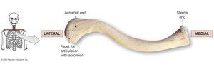
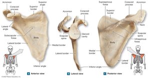
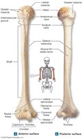
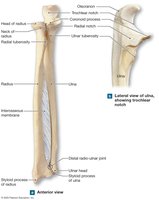
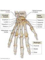
Pelvic Girdle and Lower Limb
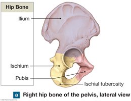
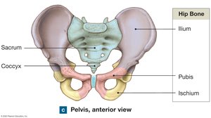
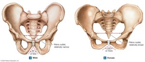
Pelvic girdle: Two coxal (hip) bones, each formed by fusion of ilium, ischium, and pubis; articulates with sacrum and femur.
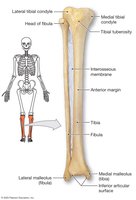
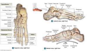
Lower limb: Femur (thigh), patella (kneecap), tibia and fibula (leg), tarsals (ankle), metatarsals (foot), phalanges (toes).
Joints (Articulations)
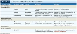
Classification of Joints
Joints are classified by structure and function:
Functional Category | Structural Category and Type | Description | Example |
|---|---|---|---|
Synarthrosis (No movement) | Fibrous (Suture, Gomphosis), Cartilaginous (Synchondrosis) | Immovable joints | Sutures of skull, teeth sockets, epiphyseal plates |
Amphiarthrosis (Little movement) | Fibrous (Syndesmosis), Cartilaginous (Symphysis) | Slightly movable joints | Distal tibia-fibula, pubic symphysis, intervertebral discs |
Diarthrosis (Free movement) | Synovial | Freely movable joints | Shoulder, elbow, knee |
Synovial Joints
Most common and freely movable joints.
Components: articular cartilage, joint capsule, synovial membrane, synovial fluid.
Accessory structures: menisci, fat pads, bursae, ligaments.
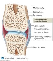
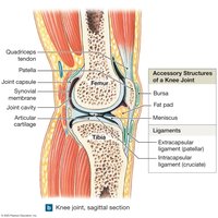
Types of Synovial Joint Movements
Gliding: Surfaces slide past each other (e.g., carpal bones).
Angular: Flexion, extension, hyperextension, abduction, adduction, circumduction.
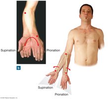
Rotation: Turning around the longitudinal axis (e.g., head, forearm pronation/supination).
Special movements: Inversion, eversion, dorsiflexion, plantar flexion, opposition, elevation, depression, lateral flexion.
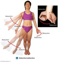
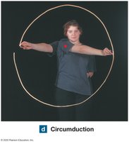
Types of Synovial Joints
Plane (gliding): Slight movement (e.g., intercarpal joints).
Hinge: Angular movement in one plane (e.g., elbow, knee).
Saddle: Angular movement in two planes (e.g., thumb joint).
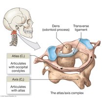
Pivot: Rotation only (e.g., atlas-axis joint).
Condylar (ellipsoidal): Angular movement in two planes (e.g., wrist).
Ball-and-socket: Movement in all planes (e.g., shoulder, hip).
Articulations of the Axial and Appendicular Skeletons
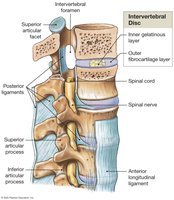
Intervertebral joints: Gliding and symphyseal joints; intervertebral discs provide cushioning.
Shoulder joint: Ball-and-socket; greatest range of motion, less stability.
Elbow joint: Hinge joint; very stable due to interlocking bones and strong ligaments.
Hip joint: Ball-and-socket; extremely stable, deep acetabulum, strong ligaments.
Knee joint: Complex hinge and plane joint; stabilized by menisci, ligaments, and tendons.
Effects of Aging on the Skeletal System
Osteopenia and Osteoporosis
Osteopenia: Age-related reduction in bone mass due to decreased osteoblast activity.
Osteoporosis: Severe bone loss, increased fracture risk; more common in postmenopausal women due to decreased estrogen.
Functional Relationships with Other Body Systems
Bones provide attachment sites for muscles (muscular system).
Bone marrow produces blood cells (cardiovascular and lymphatic systems).
Endocrine system regulates bone growth and calcium balance.
Digestive and urinary systems supply minerals for bone maintenance.
