 Back
BackChapter 7: The Skeleton – Structure and Function
Study Guide - Smart Notes
 Tailored notes based on your materials, expanded with key definitions, examples, and context.
Chapter 7: The Skeleton
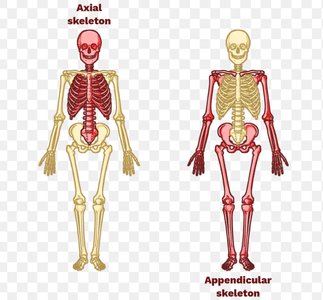
The Axial Skeleton
The axial skeleton forms the longitudinal axis of the body and is essential for protecting vital organs and supporting the head, neck, and trunk. It consists of the skull, vertebral column, and thoracic cage.
Components: Skull, vertebral column, thoracic cage (rib cage)
Functions: Protects brain, spinal cord, thoracic organs; supports head, neck, trunk
The Skull
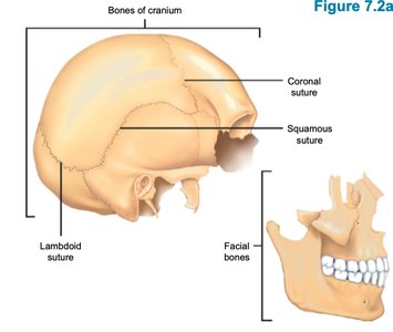
The skull is composed of cranial and facial bones, most of which are flat and joined by immovable sutures. It houses 85 openings for nerves, arteries, and veins.
Cranial Vault: Forms the superior, lateral, and posterior portions of the skull and forehead
Cranial Base: Forms the inferior aspect of the skull
Sutures: Joints between skull bones; include coronal, sagittal, lambdoid, and squamous sutures
Cranium Bones
Frontal Bone: Forms the anterior portion of the cranium; ends at supraorbital margins; contains supraorbital foramen for artery and nerve passage
Parietal Bones: Paired bones; connected by coronal, sagittal, lambdoid, and squamous sutures
Occipital Bone: Forms the posterior wall; contains foramen magnum for spinal cord passage; external occipital protuberance above foramen magnum
Temporal Bone: Includes zygomatic process and arch (cheek bones), tympanic region (external acoustic canal), and petrous region (middle and internal ear cavities)
Sphenoid Bone: Bat-shaped bone with several foramina
Ethmoid Bone: Deepest skull bone; forms roof of nasal cavity and floor of anterior cranial fossa
Facial Bones
Mandible: Largest and strongest bone; forms jaw; includes mandibular angle, alveolar process, mental foramen
Maxillary Bone: Upper jaw; holds upper teeth in alveolar processes; includes zygomatic, palatine, frontal, and infraorbital processes
Zygomatic Bones: Form cheekbones
Nasal Bones: Form bridge of nose
Lacrimal Bones: Allow for tears
Palatine Bones: Form hard palate and part of nasal cavity
Inferior Nasal Conchae: Form part of lateral nasal cavity
Hyoid Bone: Not a skull bone; movable base for tongue; attaches muscles for swallowing and speech
Vertebral Column
The vertebral column extends from the skull to the pelvis and is divided into five regions. It provides structural support and protects the spinal cord.
Regions:
Cervical (7 vertebrae)
Thoracic (12 vertebrae; 12 ribs: 6 true, 4 false, 2 floating)
Lumbar (5 vertebrae)
Sacrum (1 vertebra)
Coccyx (4 vertebrae)
Curvatures: Cervical & lumbar (concave); thoracic & sacral (convex)
Ligaments: Anterior & posterior longitudinal ligaments prevent hyperflexion & hyperextension
Intervertebral Discs: Cushion pads between vertebrae; act as shock absorbers
Nucleus Pulposus: Central, gelatinous region; provides elasticity
Anulus Fibrosus: Collagen and fibrocartilage; limits expansion
Homeostatic Imbalance: Herniated (prolapsed) disc can press on spinal cord/nerves, causing numbness/pain; treatments include exercise, massage, heat, surgery
Abnormal Curvatures:
Scoliosis: Lateral rotation (S curve); can cause breathing difficulty
Kyphosis: Dorsal thoracic curve (hunchback); seen in osteoporosis, rickets, tuberculosis
Lordosis: Lumbar curvature (swayback); seen in disease, pregnancy, obesity
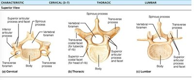
Vertebrae Structure
Body (Centrum): Main weight-bearing region
Vertebral Arch: Formed by pedicles (sides) and laminae (posterior plates)
Vertebral Foramen: Central hole for spinal cord
Vertebral Canal: Series of vertebral foramina
Intervertebral Foramina: Lateral openings for nerve passage
Processes: Spinous (posterior), transverse (lateral), superior/inferior articular (up/down)
Unique Cervical Vertebrae
C1 (Atlas): No body/spinous process; articulates with occipital condyles; allows nodding "yes"
C2 (Axis): Has body and dens; allows rotation "no"
Thoracic Vertebrae
Body: Heart-shaped
Spinous Process: Long and sharp
T1-T12: Articulate with ribs
Pectoral Girdle and Upper Limb
The pectoral girdle consists of the clavicle and scapula, connecting the upper limbs to the axial skeleton. The upper limb includes the arm, forearm, and hand.
Clavicle: Attaches upper limbs to axial skeleton
Acromioclavicular Joint: Connects clavicle and scapula
Scapula: Glenoid cavity is unstable, making shoulder dislocations common
Upper Limb: Arm, forearm, hand
