 Back
BackChapter 7: The Skeleton – Structure and Function of the Human Skeleton
Study Guide - Smart Notes
 Tailored notes based on your materials, expanded with key definitions, examples, and context.
The Skeleton
Overview of the Human Skeleton
The human skeleton is a complex framework of bones, joints, ligaments, and cartilages that provides structure, protection, and movement for the body. It is divided into two main parts: the axial skeleton and the appendicular skeleton.
Bones: Rigid organs that form the skeleton's structure.
Joints: Junctions where two or more bones meet, allowing for movement and flexibility.
Ligaments: Dense connective tissue bands that connect bones and stabilize joints.
Cartilages: Flexible connective tissues found in the nose, ear, articular surfaces of joints, costal regions, intervertebral discs, and pubic symphysis.
The Axial Skeleton
Structure and Function
The axial skeleton consists of 80 bones in three major regions: the skull, vertebral column, and thoracic cage. Its primary functions are to form the longitudinal axis of the body, support the head, neck, and trunk, and protect the brain, spinal cord, and thoracic organs.
Skull
Vertebral column
Thoracic cage
The Skull
General Features
The skull is composed of 22 bones and contains 85 named openings. It is divided into cranial bones, which enclose and protect the brain, and facial bones, which form the framework of the face and provide cavities for special senses and passageways for air and food.
Cranial bones: Protect the brain and provide attachment sites for head and neck muscles.
Facial bones: Form the face, house sensory organs, and allow for air and food passage.
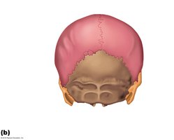
Skull Sutures
Most skull bones are flat and firmly locked together by immovable joints called sutures. Major sutures include:
Coronal suture
Squamous suture
Lambdoid suture
Sagittal suture
Cranium Geography
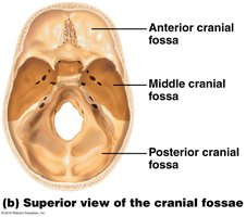
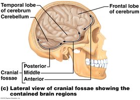
The cranium is divided into several regions and fossae that accommodate different parts of the brain.
Anterior cranial fossa: Houses the frontal lobes of the cerebrum.
Middle cranial fossa: Accommodates the temporal lobes.
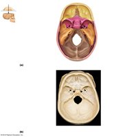
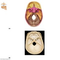
Posterior cranial fossa: Contains the cerebellum.
Major Cranial Bones and Landmarks
Frontal Bone: Forms the forehead; contains the supraorbital foramen.
Parietal Bones: Form the superior and lateral aspects of the skull.
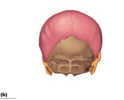
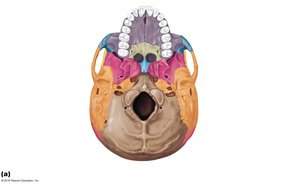
Occipital Bone: Forms the posterior part of the skull; contains the foramen magnum (where the spinal cord connects to the brain) and occipital condyles (articulate with the first vertebra, allowing head nodding).
Temporal Bones: Contain the zygomatic process (cheek), external auditory meatus (ear canal), mastoid process (neck muscle anchor), and styloid process (neck and tongue attachment).
Sphenoid Bone: Butterfly-shaped bone at the base of the skull; contains the sella turcica (pituitary gland sits here).
Ethmoid Bone: Located between the nasal cavity and orbits; features the crista galli (secures brain) and cribriform plate (roof of nasal cavity).
Facial Bones
The facial skeleton consists of 14 bones that form the structure of the face, including the mandible (lower jaw), maxillae (upper jaw), zygomatic bones (cheekbones), nasal bones, lacrimal bones, palatine bones, vomer, and inferior nasal conchae.
Mandible: Only moveable bone in the skull; forms the temporomandibular joint (TMJ).
Maxillae: Form the upper jaw and hard palate (roof of the mouth).
Zygomatic Bones: Form the prominence of the cheeks.
Nasal Bones: Form the bridge of the nose.
Lacrimal Bones: Small bones forming part of the medial orbit wall.
Palatine Bones: Complete the posterior part of the hard palate.
Vomer: Forms part of the nasal septum.
Inferior Nasal Conchae: Form part of the lateral walls of the nasal cavity.
Hyoid Bone
The hyoid bone is a unique, U-shaped bone in the neck that does not articulate directly with any other bone. It serves as an attachment site for tongue and neck muscles.
Orbits and Sinuses
Orbits: Bony cavities that house the eyes.
Sinuses: Mucosa-lined, air-filled spaces that lighten the skull and enhance voice resonance.
Vertebral Column
Structure and Function
The vertebral column consists of 26 bones in adults (33 in infants and fetuses). It disperses the weight of the trunk to the lower limbs, protects the spinal cord, and provides attachment points for ribs and back/neck muscles. The column is supported by ligaments and muscles.
Intervertebral Discs
Intervertebral discs are pads of fibrocartilage between vertebrae that absorb shock and allow flexibility. Each disc has:
Nucleus pulposus: Central, gel-like core that provides elasticity and compressibility.
Anulus fibrosus: Surrounding ring of collagen and fibrocartilage that limits expansion and holds the nucleus pulposus in place. Rupture can result in a herniated disc.
Vertebral Structure
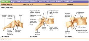
Each vertebra has a body, vertebral arch, and several processes for muscle and ligament attachment. The intervertebral foramen allows passage of spinal nerves.
Regional Vertebrae
Cervical (C1–C7): Smallest vertebrae; support the head and allow for a wide range of motion.
Thoracic (T1–T12): Articulate with ribs; have longer spinous processes.
Lumbar (L1–L5): Largest vertebrae; support the entire body weight.
Sacrum (1) & Coccyx (1): Sacrum forms the posterior wall of the pelvis; coccyx is the tailbone.
Thoracic Cage
Structure and Function
The thoracic cage consists of the thoracic vertebrae, ribs, sternum, and costal cartilages. It protects thoracic organs and supports the shoulder girdles and upper limbs.
Sternum: Composed of the manubrium (articulates with clavicles and first two pairs of ribs), body (articulates with costal cartilages), and xiphoid process (attachment for abdominal muscles).
Ribs (24): 12 pairs classified as true (1–7), false (8–10), and floating (11–12).
The Appendicular Skeleton
Structure and Function
The appendicular skeleton consists of 126 bones and is primarily responsible for movement. It includes the pectoral girdles (shoulder), pelvic girdle (hip), and the bones of the upper and lower limbs.
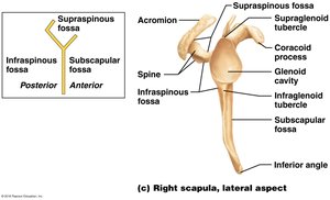
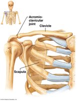
Pectoral girdle: Clavicles and scapulae attach the upper limbs to the trunk.
Pelvic girdle: Sacrum and os coxae attach the lower limbs to the trunk and support pelvic organs.
Upper Limb
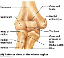
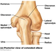
Humerus: The single bone of the upper arm.
Ulna and Radius: The two bones of the forearm; the ulna is medial, and the radius is lateral in anatomical position.
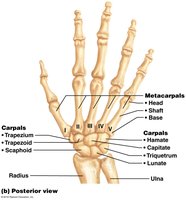
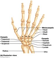
Hand: Composed of carpals (8), metacarpals (5), and phalanges (14).
Pelvic Girdle and Lower Limb
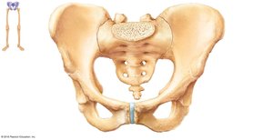
The pelvic girdle attaches the lower limbs to the axial skeleton and transmits the weight of the upper body to the lower limbs. It is composed of the sacrum and two os coxae (hip bones), each formed by the fusion of the ilium, ischium, and pubis.
Ilium: Superior region; iliac crest is the site where hands rest on hips.
Ischium: Inferior, posterior part; ischial tuberosity supports body weight when sitting.
Pubis: Anterior part; forms the pubic arch and obturator foramen.
Sexual Dimorphism of the Pelvis
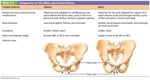
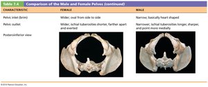
The male and female pelvis differ in structure to accommodate childbirth in females. Key differences include the shape of the pelvic inlet, the angle of the pubic arch, and the overall robustness of the bones.
Characteristic | Female | Male |
|---|---|---|
General Structure | Tilted forward, adapted for childbirth | Tilted less far forward, adapted for support |
Bone Thickness | Lighter, thinner, smoother | Heavier, thicker, more prominent markings |
Pelvic Inlet | Wider, oval | Narrow, heart-shaped |
Pubic Arch | Broader (80–90°) | More acute (50–60°) |
Lower Limb
Femur: Thigh bone; largest and strongest bone in the body.
Patella: Kneecap; protects the knee joint.
Tibia and Fibula: Bones of the lower leg; tibia is medial and weight-bearing, fibula is lateral and stabilizes the ankle.
Foot: Composed of tarsals (7), metatarsals (5), and phalanges (14).
