 Back
BackChapter 7: The Skeleton – Structure, Function, and Developmental Aspects
Study Guide - Smart Notes
 Tailored notes based on your materials, expanded with key definitions, examples, and context.
The Skeletal System Overview
Composition and Divisions
The skeletal system is a structural framework for the human body, composed primarily of bone, with cartilage in select areas and ligaments connecting bones and reinforcing joints. It accounts for approximately 20% of body mass and is divided into two major regions: the axial skeleton and the appendicular skeleton.
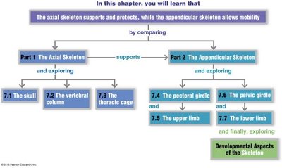
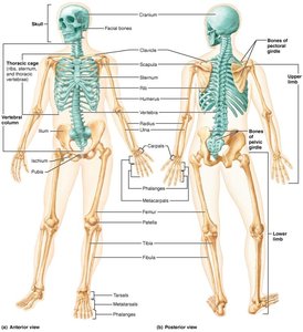
Axial skeleton: Supports and protects vital organs; forms the longitudinal axis of the body.
Appendicular skeleton: Facilitates movement; includes limbs and girdles.
Axial Skeleton
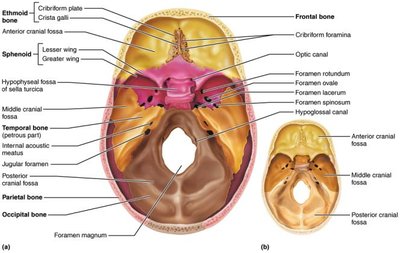
Skull
The skull is the most complex bony structure, formed by two sets of bones: cranial bones (8) and facial bones (14). Cranial bones enclose the brain and provide muscle attachment sites, while facial bones form the framework of the face, secure teeth, and anchor muscles for expression.
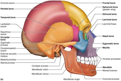
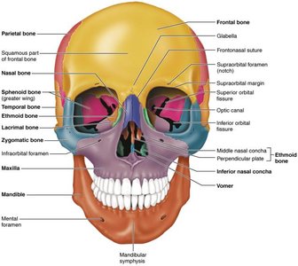
Cranial bones: Frontal, parietal, occipital, temporal, sphenoid, ethmoid.
Facial bones: Mandible, maxillae, zygomatic, nasal, lacrimal, palatine, vomer, inferior nasal conchae.
Sutures: Immovable joints between cranial bones (coronal, sagittal, squamosal, lambdoidal).
Frontal Bone
Forms the anterior cranium and superior orbit; contains the glabella and frontal sinuses.
Parietal and Occipital Bones
Parietal bones form the superior and lateral cranium; occipital bone forms the posterior wall and base, featuring the foramen magnum and occipital condyles.
Temporal Bone
Forms the inferior and lateral skull; houses auditory and vestibular mechanisms, mastoid and styloid processes.
Sphenoid and Ethmoid Bones
Sphenoid is a keystone bone, articulating with all cranial bones; ethmoid is the deepest cranial bone, forming the roof of the nasal cavity and part of the nasal septum.
Facial Skeleton
Mandible is the largest, strongest facial bone; maxillae form the upper jaw; zygomatic bones form cheekbones; nasal, lacrimal, palatine, vomer, and inferior nasal conchae complete the facial skeleton. The hyoid bone is unique, not articulating with other bones, serving as a base for the tongue and attachment for swallowing and speech muscles.
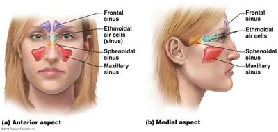
Paranasal Sinuses
Sinuses are mucosa-lined, air-filled spaces in frontal, sphenoid, ethmoid, and maxillary bones. They lighten the skull, warm and humidify air, and enhance voice resonance.
Vertebral Column
The vertebral column, or spine, transmits trunk weight to lower limbs, protects the spinal cord, and provides attachment points for ribs and muscles. It consists of 26 vertebrae: cervical (7), thoracic (12), lumbar (5), sacrum (1, fused from 5), and coccyx (1, fused from 4).
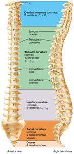
Curvatures: Cervical, thoracic, lumbar, sacral – increase flexibility and functionality.
Intervertebral discs: Pads between vertebrae, acting as shock absorbers; composed of nucleus pulposus (gelatinous center) and anulus fibrosus (fibrocartilage outer ring).
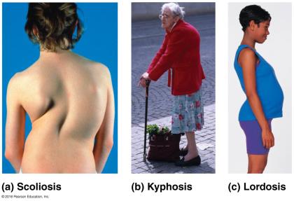
Abnormal Spinal Curvatures
Scoliosis: Lateral rotation, often in thoracic region.
Kyphosis: Dorsal thoracic curvature (hunchback).
Lordosis: Stressed lumbar curvature (swayback).
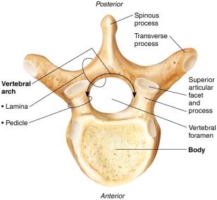
General Structure of Vertebrae
Each vertebra has a body, vertebral arch, and seven processes: spinous, transverse (2), superior articular (2), inferior articular (2).
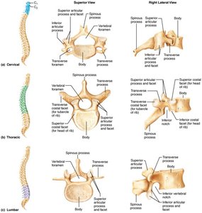
Cervical Vertebrae
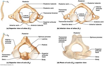
C1 (atlas) and C2 (axis) are specialized for head movement; C3–C7 are small, light, with transverse foramina for artery passage.
Thoracic and Lumbar Vertebrae
Thoracic vertebrae articulate with ribs; lumbar vertebrae are massive, supporting most body stress.
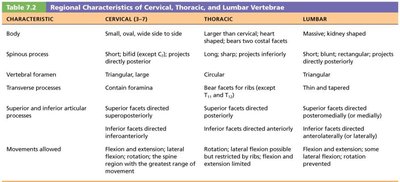
Characteristic | Cervical (C3–C7) | Thoracic | Lumbar |
|---|---|---|---|
Body | Small, oval, wide side to side | Larger than cervical, heart-shaped | Massive, kidney shaped |
Spinous process | Short, bifid (except C7) | Long, sharp, projects inferiorly | Short, blunt, rectangular |
Vertebral foramen | Triangular, large | Circular | Triangular |
Transverse processes | Contain foramina | Bear facets for ribs | Thin and tapered |
Movements allowed | Flexion, extension, lateral flexion, rotation | Rotation, lateral flexion possible | Flexion and extension |
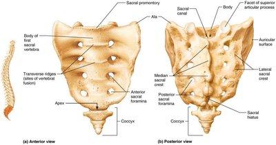
Sacrum and Coccyx
Sacrum is a triangular bone formed from five fused vertebrae, articulating with the hip bones; coccyx is formed from four fused vertebrae.
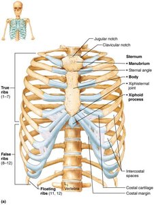
Thoracic Cage (Rib Cage)
The thoracic cage consists of thoracic vertebrae, sternum, ribs, and costal cartilages. It protects vital organs, supports shoulder girdles and upper limbs, and provides muscle attachment sites.
Sternum: Manubrium, body, xiphoid process.
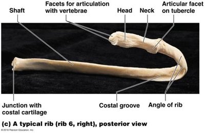
Ribs: 12 pairs – true (1–7), false (8–10), floating (11–12).
Main parts of a rib: Shaft, head, neck, tubercle, costal groove.
Appendicular Skeleton
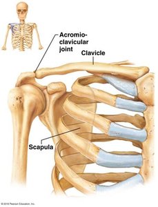
Pectoral Girdle (Shoulder Girdle)
The pectoral girdle consists of clavicles and scapulae, attaching upper limbs to the axial skeleton and providing muscle attachment sites.
Upper Limb
Each upper limb contains 30 bones: humerus, radius, ulna, carpals (8), metacarpals (5), phalanges (14).
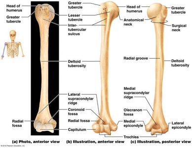
Humerus: Articulates with scapula, radius, and ulna; features include head, neck, tubercles, deltoid tuberosity, trochlea, capitulum, epicondyles, and fossae.
Ulna: Medial forearm bone, forms elbow joint; features include olecranon, coronoid process, trochlear notch, radial notch, head, styloid process.
Radius: Lateral forearm bone; features include head, radial tuberosity, ulnar notch, styloid process.

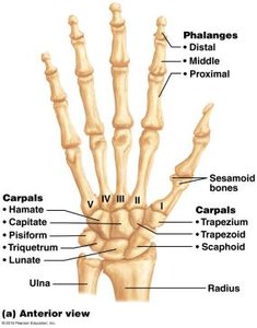
Hand
Carpals are arranged in two rows; metacarpals form the palm; phalanges form fingers. The carpal tunnel is a ligamentous tunnel for the median nerve and tendons.
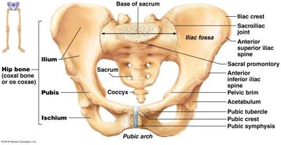
Pelvic Girdle
The pelvic girdle is formed by two coxal bones, attaching lower limbs to the axial skeleton and supporting pelvic organs. It is more stable but less mobile than the shoulder joint.
Coxal bones: Ilium (superior), ischium (inferior/posterior), pubis (inferior/anterior).
Pubic symphysis: Joins pubic bones.
Acetabulum: Deep socket for femur head.
Pubic arch: Angle between pubic bones, wider in females for childbearing.
Lower Limb
The lower limb includes the femur, patella, tibia, fibula, tarsals, metatarsals, and phalanges.
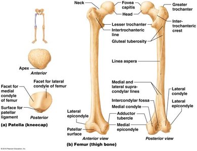
Femur: Largest, strongest bone; articulates with acetabulum, tibia, and patella; features include fovea capitis, trochanters, linea aspera, condyles, epicondyles, patellar surface, intercondylar fossa.
Patella: Sesamoid bone protecting knee joint.
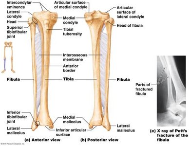
Tibia: Medial leg bone, weight-bearing; features include condyles, tuberosity, medial malleolus.
Fibula: Lateral leg bone, not weight-bearing; features include lateral malleolus.
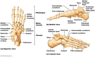
Foot
The foot includes tarsal bones (7), metatarsals (5), and phalanges (14). The arches of the foot (lateral longitudinal, medial longitudinal, transverse) are maintained by bones, ligaments, and tendons, allowing weight bearing. Fallen arches (flat feet) result from stress on tendons and ligaments.
Developmental Aspects of the Skeleton
Fontanels
Fontanels are membrane-filled spaces between cranial bones in infants, allowing skull compression during birth. They ossify over time, with six major fontanels: frontal (anterior), occipital (posterior), sphenoid (anterolateral, 2), mastoid (posterolateral, 2).
Infant and Aging Skeleton
Infant skull has more bones, connected by fontanelles.
Primary curvatures (thorax, sacrum) are convex at birth; secondary curvatures (cervical, lumbar) develop as child grows.
With age, intervertebral discs thin, costal cartilages may ossify, bones lose mass, and fracture risk increases.
Key Terms and Concepts
Bone: Rigid organ forming the skeleton.
Cartilage: Flexible connective tissue in joints.
Ligament: Connects bones, reinforces joints.
Suture: Immovable joint between skull bones.
Fontanel: Soft spot in infant skull.
Condyle: Rounded articular projection.
Process: Projection for muscle/ligament attachment.
Summary Table: Major Bones of the Skeleton
Region | Main Bones |
|---|---|
Skull | Frontal, parietal, occipital, temporal, sphenoid, ethmoid, mandible, maxilla, zygomatic, nasal, lacrimal, palatine, vomer, inferior nasal conchae |
Vertebral Column | Cervical, thoracic, lumbar vertebrae, sacrum, coccyx |
Thoracic Cage | Sternum, ribs, costal cartilages |
Pectoral Girdle | Clavicle, scapula |
Upper Limb | Humerus, radius, ulna, carpals, metacarpals, phalanges |
Pelvic Girdle | Ilium, ischium, pubis |
Lower Limb | Femur, patella, tibia, fibula, tarsals, metatarsals, phalanges |
Equations and Formulas
Bone Mass Percentage:
Additional info:
Fontanels allow for rapid brain growth in infancy.
Differences in male and female pelvis are adaptations for childbirth.
Carpal tunnel syndrome is a clinical condition relevant to the anatomy of the wrist.
