 Back
Chapter 8: Articulations (Joints) – ANP College Study Guide
Study Guide - Smart Notes
 Tailored notes based on your materials, expanded with key definitions, examples, and context.
Joints and Articulations
Definition and Function
Joints, also known as articulations, are the locations where two or more bones meet. They may also occur between bone and cartilage or between bone and teeth. The primary functions of joints are to provide both stability and mobility to the skeleton. Most joints represent a compromise between these two requirements, with some joints being highly mobile but less stable, and others being very stable but less mobile.
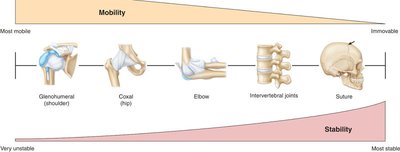
Stability: Prevents excessive movement and protects internal structures.
Mobility: Allows for movement necessary for daily activities and locomotion.
Classification of Joints
Functional Classification
Joints are classified functionally based on the degree of movement they permit:
Synarthrosis: Immovable joints (e.g., sutures of the skull).
Amphiarthrosis: Slightly movable joints (e.g., intervertebral discs).
Diarthrosis: Freely movable joints (e.g., shoulder, hip).
Structural Classification
Joints are also classified structurally based on how the bones are held together:
Fibrous Joints: Bones joined by dense connective tissue; mostly immovable or slightly movable.
Cartilaginous Joints: Bones joined by cartilage; allow limited movement.
Synovial Joints: Bones separated by a fluid-filled cavity; freely movable.
Fibrous Joints
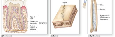
Fibrous joints include sutures, syndesmoses, and gomphoses. These joints are typically immovable or allow very limited movement.
Cartilaginous Joints
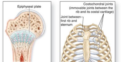
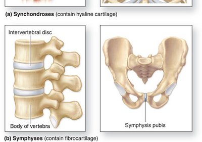
Cartilaginous joints are classified as either synchondroses (contain hyaline cartilage) or symphyses (contain fibrocartilage).
Synovial Joints
Synovial joints are the most common and most movable type of joint in the body. They are characterized by a joint cavity filled with synovial fluid.
Types of Synovial Joints
Six Subtypes of Synovial Joints
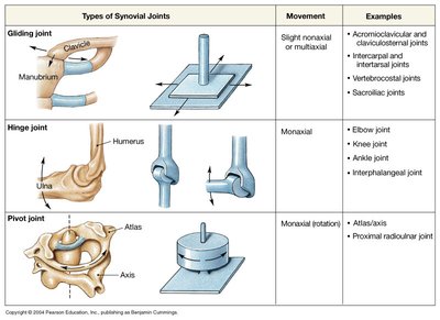
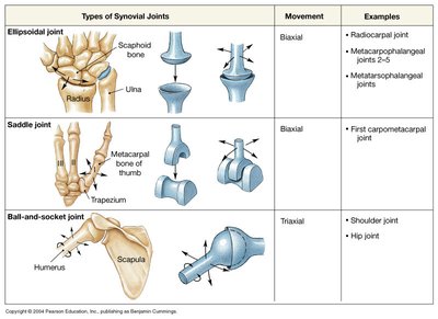
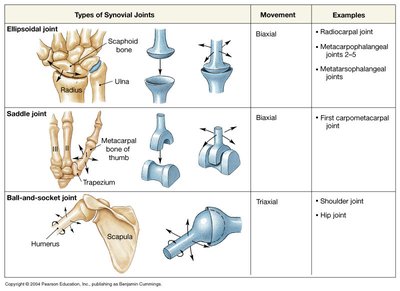
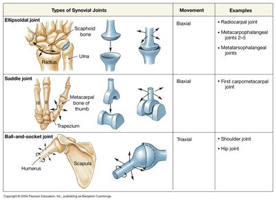
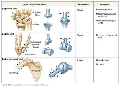
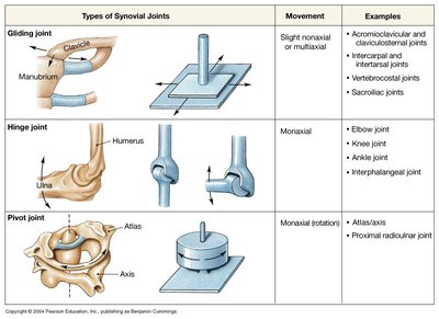
Synovial joints are classified into six types based on the shapes of their articulating surfaces and the movements they allow:
Planar (Gliding) Joint: Allows sliding or gliding movements.
Hinge Joint: Permits flexion and extension (e.g., elbow, knee).
Pivot Joint: Allows rotational movement (e.g., atlas/axis).
Condyloid (Ellipsoid) Joint: Permits movement in two planes (e.g., wrist).
Saddle Joint: Allows movement in two planes, with greater freedom than condyloid (e.g., thumb).
Ball-and-Socket Joint: Allows movement in multiple axes and planes (e.g., shoulder, hip).
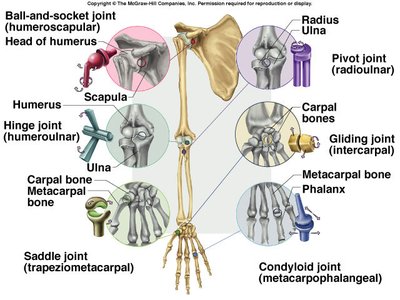
Synovial Joint Types Table
Type | Movement | Examples |
|---|---|---|
Gliding (Planar) | Slight monaxial or multiaxial | Acromioclavicular, intercarpal, vertebrocostal |
Hinge | Monaxial (flexion/extension) | Elbow, knee, ankle, interphalangeal |
Pivot | Monaxial (rotation) | Atlas/axis, proximal radioulnar |
Ellipsoid (Condyloid) | Biaxial | Radiocarpal, metacarpophalangeal |
Saddle | Biaxial | First carpometacarpal (thumb) |
Ball-and-Socket | Triaxial | Shoulder, hip |
Action Terms: Movements at Joints
Basic Movements
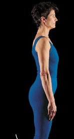
Joints allow a variety of movements, each with specific terminology:
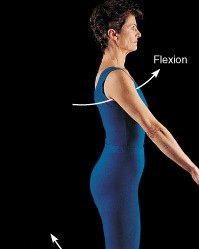
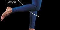
Flexion: Decreases the angle between two bones.
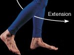
Extension: Increases the angle between two bones.
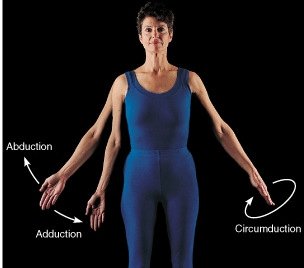
Abduction: Movement away from the midline.
Adduction: Movement toward the midline.
Circumduction: Circular movement combining flexion, extension, abduction, and adduction.
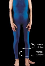
Rotation: Movement around a longitudinal axis (medial/lateral).
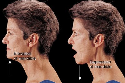
Elevation/Depression: Raising or lowering a body part.
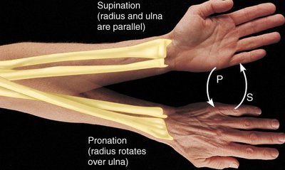
Supination/Pronation: Rotational movement of the forearm.
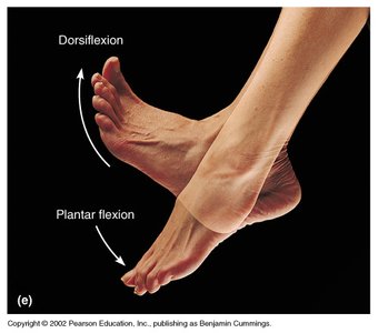
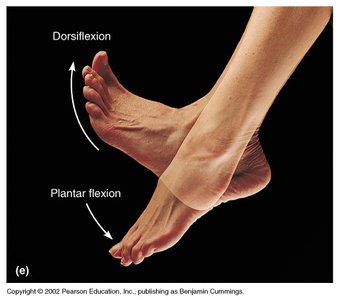
Dorsiflexion/Plantarflexion: Movements at the ankle.
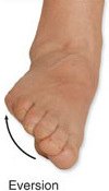
Inversion/Eversion: Movements of the sole of the foot.
Protraction/Retraction: Moving a body part forward or backward.

Synovial Joint Structures
General Anatomy of Synovial Joints
Synovial joints have a unique structure that allows for free movement:
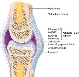
Joint cavity: Space between articulating bones filled with synovial fluid.
Synovial fluid: Lubricates and nourishes the joint.
Articular cartilage: Covers bone surfaces, reducing friction and absorbing shock.
Articular capsule: Encloses the joint cavity; consists of fibrous and synovial layers.
Synovial membrane: Produces synovial fluid.
Ligaments: Strengthen and stabilize the joint.
Accessory Structures
Bursa: Fluid-filled sacs that reduce friction between tissues.
Tendon sheath: Tubular bursae surrounding tendons.
Tendons: Connect muscle to bone, contributing to joint stability.
Ligaments: Connect bone to bone, reinforcing the joint.
Menisci: Pads of fibrocartilage that improve fit and absorb shock.
The Knee Joint
Structures of the Knee Joint
The knee is a complex synovial joint with several important structures:
Anterior cruciate ligament (ACL): Prevents anterior displacement of the tibia.
Posterior cruciate ligament (PCL): Prevents posterior displacement of the tibia.
Lateral collateral ligament (LCL): Stabilizes the lateral aspect of the knee.
Medial collateral ligament (MCL): Stabilizes the medial aspect of the knee.
Lateral and medial meniscus: C-shaped cartilage pads that cushion and stabilize the joint.
Joint Disorders
Common Disorders
Sprain: Stretching or tearing of ligaments.
Dislocation: Displacement of bones at a joint.
Osteoarthritis: Most common form; chronic, slow-progressive, irreversible. Results from aging, with softening and degeneration of cartilage, development of bony spurs, pain, and restricted movement.
Rheumatoid Arthritis: Chronic, rapid onset, autoimmune disorder affecting 2.5% of adults. Characterized by variable flare-ups; inflammation controlled by steroids and aspirin.
Comparison Table: Osteoarthritis vs. Rheumatoid Arthritis
Feature | Osteoarthritis | Rheumatoid Arthritis |
|---|---|---|
Onset | Slow, progressive | Rapid, variable |
Cause | Aging, wear-and-tear | Autoimmune |
Cartilage | Degeneration, bony spurs | Inflammation, erosion |
Population | Older adults | 2.5% of adults |
Treatment | Pain management | Steroids, aspirin |
Note: Osteoarthritis is not related to osteoporosis.
Summary
Joints are essential for movement and stability in the human body. Understanding their classification, structure, and function is fundamental for anatomy students. Synovial joints, with their complex anatomy and wide range of movements, are particularly important. Awareness of common joint disorders aids in clinical understanding and application.
