 Back
BackChapter 8: Joints (Articulations) – Structure, Function, and Clinical Aspects
Study Guide - Smart Notes
 Tailored notes based on your materials, expanded with key definitions, examples, and context.
Joints (Articulations)
Introduction to Joints
Joints, or articulations, are sites where two or more bones meet. They are essential for providing the skeleton with mobility and holding the skeleton together. Understanding joints is crucial for diagnosing and treating musculoskeletal injuries and disorders.
Classification of Joints
Structural Classification
Joints are classified structurally based on the material binding the bones and the presence or absence of a joint cavity:
Fibrous Joints: Bones joined by dense fibrous connective tissue; no joint cavity.
Cartilaginous Joints: Bones united by cartilage; no joint cavity.
Synovial Joints: Bones separated by a fluid-filled joint cavity.
Functional Classification
Joints are also classified by the degree of movement they allow:
Synarthroses: Immovable joints
Amphiarthroses: Slightly movable joints
Diarthroses: Freely movable joints
Fibrous Joints
General Features
Fibrous joints are connected by dense fibrous connective tissue and lack a joint cavity. Most are immovable, but the degree of movement depends on the length of the connective tissue fibers.
Types of Fibrous Joints
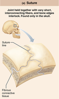
Sutures: Rigid, interlocking joints found only in the skull. They allow for growth during youth and ossify in adulthood to form synostoses (immovable joints).
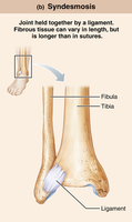
Syndesmoses: Bones connected by ligaments. The amount of movement depends on fiber length. Example: the distal tibiofibular joint (little movement) and the interosseous membrane between the radius and ulna (more movement).
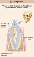
Gomphoses: Peg-in-socket joints, such as teeth in their alveolar sockets. The periodontal ligament holds the tooth in place.
Cartilaginous Joints
General Features
Cartilaginous joints unite bones with cartilage and lack a joint cavity. They are not highly movable.
Types of Cartilaginous Joints
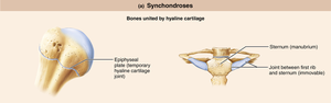
Synchondroses: Bones united by a bar or plate of hyaline cartilage. Most are immovable (synarthrotic). Examples include the epiphyseal plates in growing children and the joint between the first rib and the manubrium of the sternum.
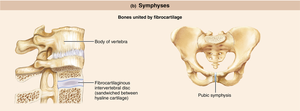
Symphyses: Bones united by fibrocartilage, with hyaline cartilage present as articular cartilage on bony surfaces. These joints are strong and slightly movable (amphiarthrotic). Examples include intervertebral joints and the pubic symphysis.
Synovial Joints
General Structure
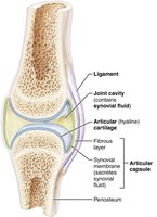
Synovial joints are characterized by the presence of a fluid-filled joint cavity and are all diarthrotic (freely movable). They are the most common type of joint in the limbs.
Articular cartilage: Hyaline cartilage covering bone ends, preventing crushing of bone ends.
Joint (synovial) cavity: Small, fluid-filled space unique to synovial joints.
Articular (joint) capsule: Two layers—an external fibrous layer (dense irregular connective tissue) and an inner synovial membrane (loose connective tissue that produces synovial fluid).
Synovial fluid: Viscous, slippery filtrate that lubricates and nourishes articular cartilage; contains phagocytic cells.
Reinforcing ligaments: Capsular, extracapsular, and intracapsular ligaments stabilize the joint.
Nerves and blood vessels: Nerves detect pain and monitor joint position; capillaries supply filtrate for synovial fluid.
Associated Structures
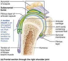
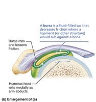
Bursae: Fluid-filled sacs that reduce friction where ligaments, muscles, skin, tendons, or bones rub together.
Tendon sheaths: Elongated bursae that wrap around tendons subjected to friction.
Factors Influencing Stability
Shape of articular surfaces: Shallow surfaces are less stable than ball-and-socket joints.
Ligament number and location: More ligaments generally increase joint strength.
Muscle tone: The most important factor; keeps tendons taut and stabilizes joints, especially in the shoulder and knee.
Movements Allowed by Synovial Joints
Types of Movement
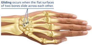
Gliding: Flat bone surfaces slide over each other. Examples: intercarpal and intertarsal joints.
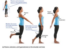
Angular Movements: Change the angle between bones.
Flexion: Decreases the angle of the joint.
Extension: Increases the angle of the joint.
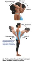
Hyperextension: Extension beyond the anatomical position.
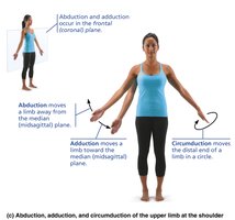
Abduction: Movement away from the midline.
Adduction: Movement toward the midline.
Circumduction: Limb describes a cone in space, combining flexion, abduction, extension, and adduction.
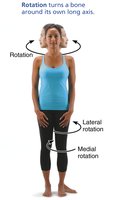
Rotation: Turning a bone around its own long axis. Medial rotation is toward the midline; lateral rotation is away from the midline. Examples: rotation of the head, humerus, and femur.
Special Movements
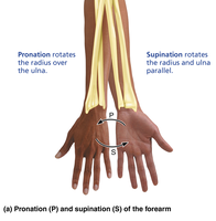
Supination and Pronation: Rotation of the radius and ulna. Supination turns the palm anteriorly; pronation turns it posteriorly.
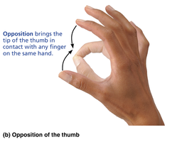
Opposition: Movement of the thumb to touch the tips of other fingers.
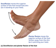
Dorsiflexion and Plantar Flexion: Dorsiflexion lifts the foot toward the shin; plantar flexion points the toes.
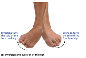
Inversion and Eversion: Inversion turns the sole of the foot medially; eversion turns it laterally.
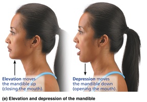
Elevation and Depression: Elevation lifts a body part superiorly (e.g., closing the mouth); depression lowers it (e.g., opening the mouth).
Protraction and Retraction: Protraction moves a body part anteriorly; retraction moves it posteriorly.
Summary Table: Types of Joints
Structural Type | Functional Type | Mobility | Example |
|---|---|---|---|
Fibrous: Suture | Synarthrosis | Immovable | Skull suture |
Fibrous: Syndesmosis | Amphiarthrosis | Slightly movable | Distal tibiofibular joint |
Fibrous: Gomphosis | Synarthrosis | Immovable | Tooth in socket |
Cartilaginous: Synchondrosis | Synarthrosis | Immovable | Epiphyseal plate |
Cartilaginous: Symphysis | Amphiarthrosis | Slightly movable | Pubic symphysis |
Synovial | Diarthrosis | Freely movable | Shoulder, knee |
Clinical Aspects of Joints
Common Joint Injuries
Cartilage Tears: Caused by compression and shear stress; often require arthroscopic surgery.
Sprains: Ligaments are stretched or torn; healing is slow due to poor blood supply.
Dislocations (Luxations): Bones are forced out of alignment; often accompanied by sprains and inflammation.
Inflammatory and Degenerative Conditions
Bursitis: Inflammation of a bursa, usually due to trauma or friction.
Tendonitis: Inflammation of tendon sheaths, typically from overuse.
Arthritis: Over 100 types; most common are osteoarthritis (degenerative), rheumatoid arthritis (autoimmune), and gouty arthritis (uric acid crystal deposition).
Lyme Disease: Bacterial infection transmitted by ticks, can lead to joint pain and arthritis.
Developmental Aspects of Joints
By embryonic week 8, synovial joints resemble adult joints. Joint structure and flexibility are influenced by use and age. Aging leads to decreased flexibility and increased risk of degenerative joint diseases.
