 Back
BackChapter 8: Joints (Articulations) – Structure, Function, and Movement
Study Guide - Smart Notes
 Tailored notes based on your materials, expanded with key definitions, examples, and context.
Joints (Articulations): Structure, Function, and Classification
Introduction to Joints
Joints, or articulations, are anatomical structures where two or more bones meet. They are essential for holding the skeleton together and providing mobility. Understanding joints is crucial for diagnosing and treating musculoskeletal injuries.
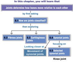
Classification of Joints
Joints are classified based on their structure and function:
Structural classification: Based on the material binding bones together and the presence or absence of a joint cavity.
Functional classification: Based on the amount of movement allowed at the joint.
Structural Types:
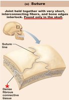
Fibrous joints: Bones joined by fibrous tissue; no joint cavity.
Cartilaginous joints: Bones joined by cartilage; no joint cavity.
Synovial joints: Bones separated by a fluid-filled joint cavity.
Functional Types:
Synarthroses: Immovable joints (mostly fibrous).
Amphiarthroses: Slightly movable joints (mostly cartilaginous).
Diarthroses: Freely movable joints (all synovial).
Fibrous Joints
General Features
Fibrous joints are connected by dense connective tissue, primarily collagen fibers. They lack a joint cavity and are mostly immovable (synarthrotic).
Types of Fibrous Joints
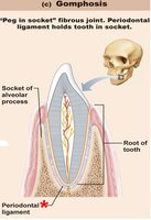
Sutures: Found only in the skull; bones are tightly bound by minimal fibrous tissue. With age, sutures may ossify and become synostoses (bony junctions).
Gomphoses: Peg-in-socket joints, such as teeth anchored in their sockets by the periodontal ligament.
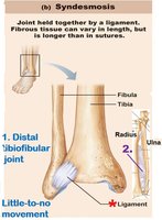
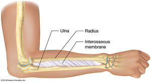
Syndesmoses: Bones connected by ligaments; movement depends on the length of the connecting fibers. Examples include the distal tibiofibular joint (little movement) and the interosseous membrane between the radius and ulna (more movement).
Cartilaginous Joints
General Features
Cartilaginous joints unite bones with cartilage and lack a joint cavity. They allow little to no movement.
Types of Cartilaginous Joints
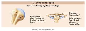
Synchondroses: Bones united by hyaline cartilage. Most are immovable (synarthrotic). Examples include the epiphyseal plates in growing bones and the joint between the first rib and the sternum.
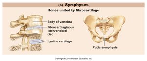
Symphyses: Bones united by fibrocartilage, which acts as a shock absorber and allows limited movement (amphiarthrotic). Examples include intervertebral discs and the pubic symphysis.
Synovial Joints
General Features
Synovial joints are characterized by a fluid-filled joint cavity and are freely movable (diarthrotic). They are the most common type of joint in the body, especially in the limbs.
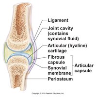
Six Distinguishing Features of Synovial Joints
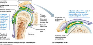
Articular cartilage: Hyaline cartilage covering bone surfaces to reduce friction.
Joint (articular) cavity: Space containing synovial fluid.
Synovial fluid: Viscous, slippery fluid that lubricates and nourishes the joint.
Articular (joint) capsule: Two-layered capsule enclosing the joint cavity (outer fibrous layer and inner synovial membrane).
Reinforcing ligaments: Strengthen and stabilize the joint (capsular, extracapsular, and intracapsular types).
Nerves and blood vessels: Supply the joint, detect pain, and monitor joint position and stretch.
Other Structural Features
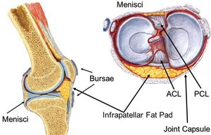
Fatty pads: Cushioning structures between the fibrous layer and bone or synovial membrane (e.g., knee and hip joints).
Articular discs (menisci): Fibrocartilage pads that improve the fit of bone ends, stabilize the joint, and reduce wear and tear.
Bursae: Fluid-filled sacs that reduce friction where ligaments, muscles, skin, or tendons rub against bone.
Tendon sheaths: Elongated bursae that wrap around tendons to decrease friction.
Stability of Synovial Joints
Three main factors influence the stability of synovial joints:
Shape of articular surfaces: Minor role in stability.
Ligament number and location: Limited role; more ligaments generally increase stability.
Muscle tone: Most important factor; keeps tendons taut and stabilizes joints, especially in the shoulder, knee, and foot arches.
Movements at Synovial Joints
General Principles
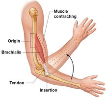
All muscles attach to bone at two points: the origin (immovable) and the insertion (movable). Muscle contraction moves the insertion toward the origin, producing movement at the joint. Movements occur along transverse, frontal, or sagittal planes.
Types of Movement
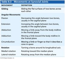
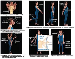
Gliding: Flat bone surfaces slide past each other (e.g., intercarpal joints).
Angular movements: Change the angle between bones. Includes flexion, extension, hyperextension, abduction, adduction, and circumduction.
Rotation: Turning a bone around its own long axis (e.g., between atlas and axis, shoulder, hip).
Movement | Definition |
|---|---|
Gliding | Sliding the flat surfaces of two bones across each other |
Flexion | Decreasing the angle between two bones, usually in the sagittal plane |
Extension | Increasing the angle between two bones, usually in the sagittal plane |
Abduction | Moving a limb away from the body midline in the frontal plane |
Adduction | Moving a limb toward the body midline in the frontal plane |
Circumduction | Moving a limb or finger so that it describes a cone in space |
Rotation | Turning a bone around its longitudinal axis |
Medial rotation | Rotating toward the median plane |
Lateral rotation | Rotating away from the median plane |
Range of Motion
Nonaxial: Slipping movements only (plane joints).
Uniaxial: Movement in one plane (hinge and pivot joints).
Biaxial: Movement in two planes (condylar and saddle joints).
Multiaxial: Movement in or around all three planes (ball-and-socket joints).
Types of Synovial Joints
Plane joint: Nonaxial, gliding movements (e.g., intercarpal joints).
Hinge joint: Uniaxial, flexion and extension (e.g., elbow, knee).
Pivot joint: Uniaxial, rotation (e.g., proximal radioulnar joint, atlantoaxial joint).
Condylar joint: Biaxial, flexion/extension and abduction/adduction (e.g., knuckle joints).
Saddle joint: Biaxial, similar to condylar but with greater movement (e.g., thumb carpometacarpal joint).
Ball-and-socket joint: Multiaxial, most freely moving (e.g., shoulder, hip).
Selected Synovial Joints: The Knee Joint
Structure and Function
The knee is the largest and most complex joint in the body, consisting of three joints in one cavity: the femoropatellar joint and the lateral and medial tibiofemoral joints. It is a modified hinge joint, allowing flexion, extension, and some rotation.
Ligaments and Stability
Capsular and extracapsular ligaments: Prevent hyperextension and stabilize the joint (e.g., tibial and fibular collateral ligaments).
Intracapsular ligaments: Cruciate ligaments (anterior and posterior) prevent anterior-posterior displacement of the tibia and femur.
Menisci: Medial and lateral fibrocartilage pads that improve fit and absorb shock.
Common Knee Injuries
The knee is vulnerable to horizontal or lateral blows, especially in sports. Common injuries involve the three C's: collateral ligaments, cruciate ligaments, and cartilages (menisci).
Summary Table: Types of Synovial Joints and Movements
Joint Type | Movement | Example |
|---|---|---|
Plane | Nonaxial (gliding) | Intercarpal joints |
Hinge | Uniaxial (flexion/extension) | Elbow, knee |
Pivot | Uniaxial (rotation) | Proximal radioulnar joint |
Condylar | Biaxial (flexion/extension, abduction/adduction) | Knuckle joints |
Saddle | Biaxial (greater movement) | Thumb carpometacarpal joint |
Ball-and-socket | Multiaxial (all movements) | Shoulder, hip |
Key Equations:
Range of motion (ROM): The degree through which a joint can move, measured in degrees of a circle.
Additional info: Understanding joint structure and function is essential for clinical assessment and treatment of joint injuries, such as sprains, dislocations, and degenerative diseases like arthritis.
