 Back
BackChapter 8: Joints (Articulations) – Structure, Function, and Clinical Aspects
Study Guide - Smart Notes
 Tailored notes based on your materials, expanded with key definitions, examples, and context.
Joints: An Overview
Definition and Function
Joints, also known as articulations, are sites where two or more bones meet. They play a crucial role in holding the skeleton together and providing mobility to the body.
Hold bones together to maintain structural integrity.
Allow for mobility by permitting various types of movement.
Classification of Joints
Joints are classified in two main ways:
Functional classification: Based on the amount of movement allowed.
Structural classification: Based on the material binding the bones and the presence or absence of a joint cavity.
Functional Classification of Joints
Synarthroses: Immovable joints (e.g., sutures of the skull).
Amphiarthroses: Slightly movable joints (e.g., intervertebral discs).
Diarthroses: Freely movable joints (e.g., most limb joints).
Structural Classification of Joints
Fibrous joints: Bones joined by dense fibrous connective tissue; generally immovable.
Cartilaginous joints: Bones joined by cartilage; immovable or slightly movable.
Synovial joints: Bones separated by a fluid-filled joint cavity; freely movable.
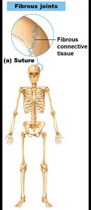
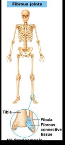
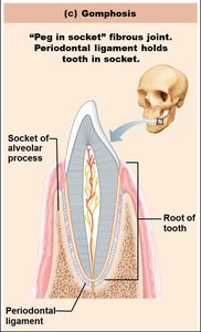
Fibrous Joints
General Features
Fibrous joints are united by dense fibrous connective tissue and lack a joint cavity. All are synarthrotic (immovable).
Sutures: Found between cranial bones; allow for growth during youth and fuse in adulthood.
Syndesmoses: Bones connected by ligaments; allow more movement than sutures but still limited (e.g., distal tibiofibular joint).
Gomphoses: Peg-in-socket joints (e.g., tooth in alveolar socket, held by periodontal ligament).
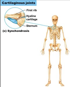
Cartilaginous Joints
General Features
Cartilaginous joints are united by cartilage and lack a joint cavity. They are either synarthrotic or amphiarthrotic.
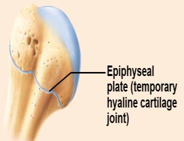
Synchondroses: Bones united by hyaline cartilage; mostly immovable (e.g., epiphyseal plates, first rib and sternum).
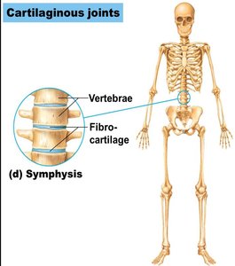
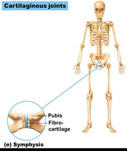
Symphyses: Bones united by fibrocartilage; slightly movable (e.g., pubic symphysis, intervertebral discs).
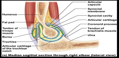
Synovial Joints
General Features
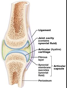
Synovial joints are the most common and movable type of joint in the body. They have a joint cavity filled with synovial fluid and are always diarthrotic (freely movable).
Articular cartilage (hyaline) covers bone ends to prevent crushing.
Joint (synovial) cavity contains synovial fluid.
Articular capsule encloses the joint cavity, consisting of an outer fibrous layer and an inner synovial membrane.
Synovial fluid lubricates and nourishes cartilage, contains phagocytes.
Reinforcing ligaments (capsular, extracapsular, intracapsular) stabilize the joint.
Nerves and blood vessels supply the joint, detect pain, and monitor position.
Additional Features in Some Synovial Joints
Fatty pads: Cushion between fibrous layer and bone or synovial membrane.
Articular discs (menisci): Fibrocartilage pads that improve fit, stabilize, and reduce wear.
Bursae: Fluid-filled sacs that reduce friction where structures rub together.
Tendon sheaths: Elongated bursae that wrap around tendons.
Stabilizing Factors for Synovial Joints
Shape of articular surfaces (minor role).
Number and location of ligaments (limited role).
Muscle tone (most important): Keeps tendons taut and stabilizes joints, especially in the shoulder, knee, and foot arches.
Key Synovial Joints: Structure and Stabilization
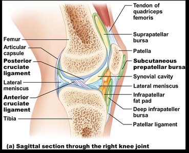
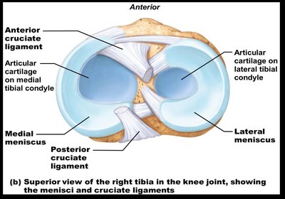
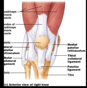
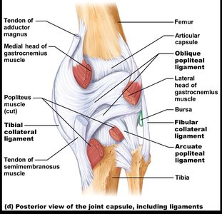
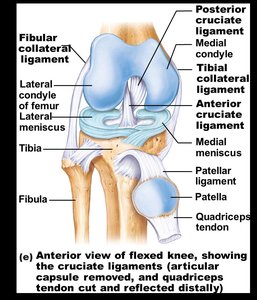
Knee Joint
The knee is the largest and most complex joint, consisting of three joints within a single cavity. It allows flexion, extension, and some rotation.
Stabilized by muscle tendons (quadriceps, semimembranosus), ligaments (patellar, collateral, cruciate), and menisci.
Contains at least 12 bursae.
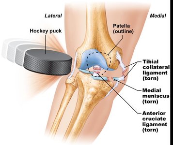
Vulnerable to injury, especially from lateral blows ("unhappy triad": torn tibial collateral ligament, medial meniscus, anterior cruciate ligament).
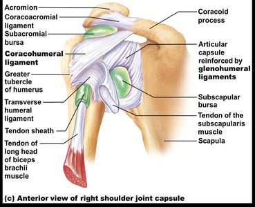
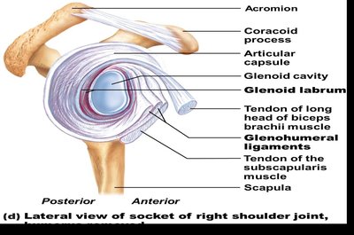
Shoulder Joint (Glenohumeral Joint)
A ball-and-socket joint with the greatest range of motion but less stability. Stabilized by the rotator cuff muscles, glenohumeral ligaments, and the coracohumeral ligament.
Rotator cuff muscles: Subscapularis, supraspinatus, infraspinatus, teres minor.
Glenoid labrum deepens the socket.
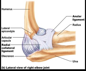
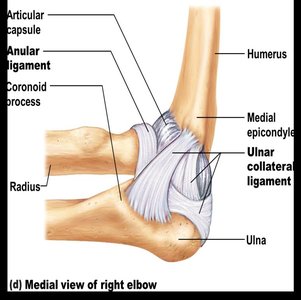
Elbow Joint
A hinge joint formed by the articulation of the humerus with the radius and ulna. Allows flexion and extension only.
Stabilized by the anular ligament (encircles the head of the radius), ulnar collateral ligament, and radial collateral ligament.
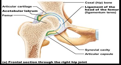
Hip (Coxal) Joint
A deep ball-and-socket joint formed by the head of the femur and the acetabulum of the pelvis. It is highly stable due to the depth of the socket and strong ligaments.
Acetabular labrum deepens the socket.
Reinforced by iliofemoral, pubofemoral, ischiofemoral ligaments, and ligamentum teres.
Temporomandibular Joint (TMJ)
The TMJ is a modified hinge joint between the mandibular condyle and the temporal bone. It allows both hinge (depression/elevation) and gliding (side-to-side) movements. It is the most easily dislocated joint in the body.
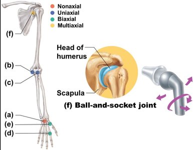
Types of Synovial Joints by Shape and Movement
Plane joint: Nonaxial, gliding movements (e.g., intercarpal joints).
Hinge joint: Uniaxial, flexion and extension (e.g., elbow).
Pivot joint: Uniaxial, rotation (e.g., proximal radioulnar joint).
Condylar joint: Biaxial, flexion/extension and abduction/adduction (e.g., knuckle joints).
Saddle joint: Biaxial, similar to condylar but with greater movement (e.g., thumb carpometacarpal joint).
Ball-and-socket joint: Multiaxial, movement in all axes (e.g., shoulder, hip).
Movements Allowed by Synovial Joints
Gliding: Flat bone surfaces slide over each other (e.g., intercarpal joints).
Angular movements: Change the angle between bones (flexion, extension, hyperextension, abduction, adduction, circumduction).
Rotation: Bone turns around its own long axis (e.g., atlas and axis, shoulder, hip).
Special movements: Include supination/pronation (forearm), dorsiflexion/plantar flexion (foot), inversion/eversion (foot), protraction/retraction (jaw), elevation/depression (mandible), opposition (thumb).
Joint Injuries and Clinical Considerations
Cartilage tears: Often involve menisci; poor healing due to avascularity.
Sprains: Ligaments stretched or torn; slow healing.
Dislocations (luxations): Bones forced out of alignment; require reduction.
Bursitis: Inflammation of a bursa, usually from trauma or friction.
Tendonitis: Inflammation of tendon sheaths, often from overuse.
Lyme disease: Bacterial infection causing joint pain and arthritis.
Types of Arthritis
Osteoarthritis (OA): Degenerative, wear-and-tear arthritis; most common in elderly.
Rheumatoid arthritis (RA): Autoimmune, chronic inflammation of joints; often bilateral.
Gouty arthritis: Uric acid crystal deposition in joints; more common in men.
Developmental Aspects of Joints
By embryonic week 8, synovial joints resemble adult joints.
Joint structure is modified by use and activity.
Aging leads to shortening and weakening of ligaments/tendons, increased risk of herniated discs, and higher prevalence of osteoarthritis.
Regular full-range-of-motion exercise helps maintain joint health.
