 Back
BackChapter 8: Joints (Articulations) – Structure, Function, and Clinical Aspects
Study Guide - Smart Notes
 Tailored notes based on your materials, expanded with key definitions, examples, and context.
Joints (Articulations)
Definition and Functions
Joints, or articulations, are sites where two or more bones meet. They are essential for providing the skeleton with mobility and for holding the skeleton together, ensuring structural integrity and movement.
Articulation: The point of contact between bones.
Functions: Mobility and stability of the skeleton.
Classification of Joints
Functional Classification: Based on the amount of movement allowed.
Synarthroses: Immovable joints
Amphiarthroses: Slightly movable joints
Diarthroses: Freely movable joints
Structural Classification: Based on the material binding bones and the presence/absence of a joint cavity.
Fibrous joints
Cartilaginous joints
Synovial joints
Fibrous Joints
General Features
Fibrous joints are connected by dense fibrous connective tissue and lack a joint cavity. Most are immovable (synarthrotic), though movement depends on the length of connective tissue fibers.
Sutures: Rigid, interlocking joints found only in the skull. Allow for growth during youth and ossify in middle age (synostoses).
Syndesmoses: Bones connected by ligaments. Movement varies with fiber length (e.g., tibiofibular joint – little movement; interosseous membrane of radius and ulna – more movement).
Gomphoses: Peg-in-socket joints (e.g., teeth in alveolar sockets), connected by the periodontal ligament.
Cartilaginous Joints
General Features
Cartilaginous joints unite bones with cartilage and lack a joint cavity. They are not highly movable.
Synchondroses: Bones united by hyaline cartilage (e.g., epiphyseal plates, first rib and manubrium). Mostly synarthrotic.
Symphyses: Bones united by fibrocartilage (e.g., intervertebral discs, pubic symphysis). Amphiarthrotic (slightly movable).
Synovial Joints
General Features
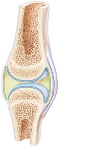
Synovial joints are characterized by bones separated by a fluid-filled joint cavity. All are diarthrotic (freely movable) and include nearly all limb joints.
Six distinguishing features:
Articular cartilage: Hyaline cartilage covering bone ends, preventing crushing.
Joint (synovial) cavity: Small, fluid-filled space.
Articular (joint) capsule: Two layers – external fibrous layer (dense irregular connective tissue) and inner synovial membrane (loose connective tissue, produces synovial fluid).
Synovial fluid: Viscous, slippery filtrate that lubricates and nourishes cartilage; contains phagocytes.
Reinforcing ligaments: Capsular (thickened fibrous layer), extracapsular (outside capsule), intracapsular (deep to capsule).
Nerves and blood vessels: Nerve fibers detect pain and monitor position; capillaries supply filtrate for synovial fluid.
Other Features of Synovial Joints
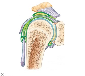
Fatty pads: Cushioning between fibrous layer and synovial membrane or bone.
Articular discs (menisci): Fibrocartilage that improves fit, stabilizes joint, and reduces wear.
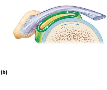
Bursae: Sacs lined with synovial membrane, reduce friction where structures rub together.
Tendon sheaths: Elongated bursae wrapped around tendons subjected to friction.
Stabilizing Factors at Synovial Joints
Shape of articular surfaces (minor role)
Ligament number and location (limited role)
Muscle tone (most important): Keeps tendons taut, especially important in shoulder, knee, and foot arches.
Summary Table: Structural and Functional Characteristics of Body Joints
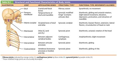
The following tables summarize the main joints, their articulating bones, structural and functional types, and movements allowed.
Illustration | Joint | Articulating Bones | Structural Type | Functional Type; Movements Allowed |
|---|---|---|---|---|
Skull | Cranial and facial bones | Fibrous; suture | Synarthrotic; no movement | |
Temporomandibular | Temporal bone of skull and mandible | Synovial; modified hinge | Diarthrotic; gliding and uniaxial rotation | |
Atlanto-occipital | Occipital bone of skull and atlas | Synovial; condyloid | Diarthrotic; biaxial: flexion, extension, lateral flexion, circumduction of head on neck | |
Atlantoaxial | Atlas (C1) and axis (C2) | Synovial; pivot | Diarthrotic; uniaxial: rotation of the head | |
Intervertebral | Between adjacent vertebral bodies | Cartilaginous; symphysis | Amphiarthrotic; slight movement | |
Intervertebral | Between articular processes | Synovial; plane | Diarthrotic; gliding | |
Costovertebral | Vertebrae and ribs | Synovial; plane | Diarthrotic; gliding of ribs |
Joint | Articulating Bones | Structural Type | Functional Type; Movements Allowed |
|---|---|---|---|
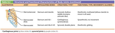
Sternoclavicular | Sternum and clavicle | Synovial; shallow saddle | Diarthrotic; multiaxial |
Sternocostal (first) | Sternum and rib 1 | Cartilaginous; synchondrosis | Synarthrotic; no movement |
Sternocostal (ribs 2–7) | Sternum and ribs II–VII | Synovial; double plane | Diarthrotic; gliding |
Joint | Articulating Bones | Structural Type | Functional Type; Movements Allowed |
|---|---|---|---|
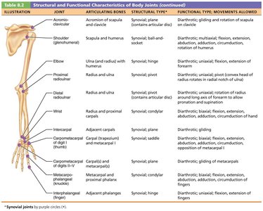
Shoulder (glenohumeral) | Scapula and humerus | Synovial; ball and socket | Diarthrotic; multiaxial |
Elbow | Ulna and radius with humerus | Synovial; hinge | Diarthrotic; uniaxial |
Proximal radioulnar | Radius and ulna | Synovial; pivot | Diarthrotic; uniaxial |
Wrist | Radius and proximal carpals | Synovial; condyloid | Diarthrotic; biaxial |
Carpometacarpal (thumb) | Carpal (trapezium) and metacarpal I | Synovial; saddle | Diarthrotic; biaxial |
Metacarpophalangeal (knuckle) | Metacarpal and proximal phalanx | Synovial; condyloid | Diarthrotic; biaxial |
Interphalangeal (finger) | Adjacent phalanges | Synovial; hinge | Diarthrotic; uniaxial |
Joint | Articulating Bones | Structural Type | Functional Type; Movements Allowed |
|---|---|---|---|
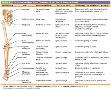
Pubic symphysis | Pubic bones | Cartilaginous; symphysis | Amphiarthrotic; slight movement |
Hip (coxal) | Hip bone and femur | Synovial; ball and socket | Diarthrotic; multiaxial |
Knee (tibiofemoral) | Femur and tibia | Synovial; modified hinge | Diarthrotic; biaxial |
Knee (femoropatellar) | Femur and patella | Synovial; plane | Diarthrotic; gliding |
Inferior tibiofibular | Tibia and fibula | Fibrous; syndesmosis | Synarthrotic; slight "give" during dorsiflexion |
Ankle | Tibia and fibula with talus | Synovial; hinge | Diarthrotic; uniaxial |
Intertarsal | Adjacent tarsals | Synovial; plane | Diarthrotic; gliding |
Tarsometatarsal | Tarsal and metatarsal | Synovial; plane | Diarthrotic; gliding |
Metatarsophalangeal | Metatarsal and proximal phalanx | Synovial; condyloid | Diarthrotic; biaxial |
Interphalangeal (toe) | Adjacent phalanges | Synovial; hinge | Diarthrotic; uniaxial |
Movements at Synovial Joints
General Principles
Muscles attach to bones at two points: origin (immovable) and insertion (movable).
Muscle contraction moves the insertion toward the origin.
Movements occur along transverse, frontal, or sagittal planes.
Range of Motion
Nonaxial: Slipping movements only
Uniaxial: Movement in one plane
Biaxial: Movement in two planes
Multiaxial: Movement in or around all three planes
Types of Movements
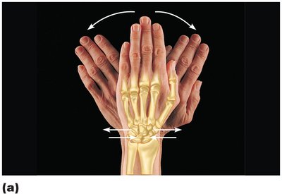
Gliding: One flat bone surface glides over another (e.g., intercarpal joints, intertarsal joints).
Angular Movements: Change the angle between bones.
Flexion: Decreases the angle of the joint
Extension: Increases the angle of the joint
Hyperextension: Movement beyond anatomical position
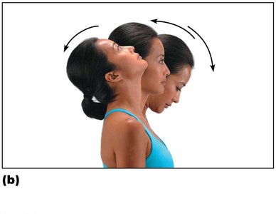
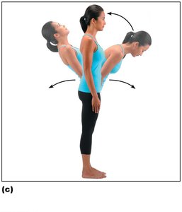
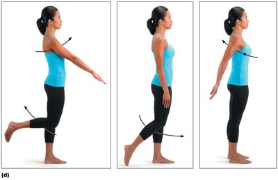
Movements along the frontal plane:
Abduction: Movement away from the midline
Adduction: Movement toward the midline
Circumduction: Limb describes a cone in space (combines flexion, abduction, extension, and adduction)
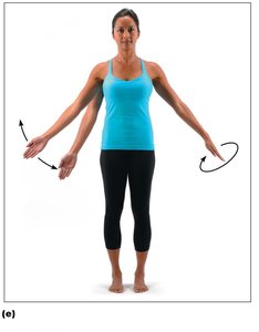
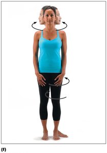
Rotation: Turning of a bone around its own long axis (e.g., between C1 and C2, rotation of humerus and femur).
Special Movements at Synovial Joints
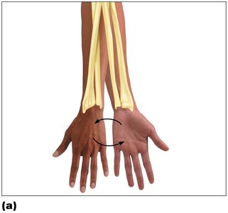
Supination and Pronation: Rotation of the radius and ulna (forearm).
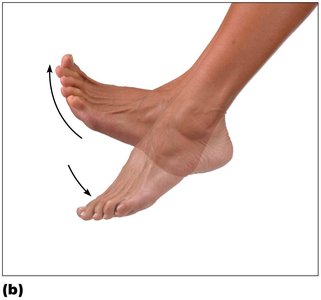
Dorsiflexion and Plantar Flexion: Upward and downward movement of the foot at the ankle.
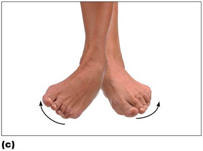
Inversion and Eversion: Turning the sole of the foot medially or laterally.
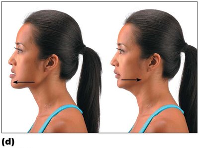
Protraction and Retraction: Anterior and posterior movement (e.g., mandible).
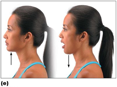
Elevation and Depression: Lifting or lowering a body part (e.g., mandible).
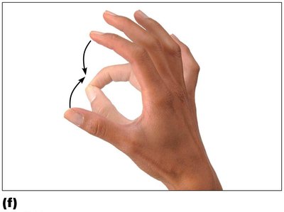
Opposition: Movement of the thumb to touch the tips of other fingers.
Types of Synovial Joints
Synovial joints are classified by the shape of their articular surfaces and the movements they allow:
Plane: Nonaxial, gliding (e.g., intercarpal joints)
Hinge: Uniaxial, flexion and extension (e.g., elbow)
Pivot: Uniaxial, rotation (e.g., proximal radioulnar joint)
Condylar: Biaxial, flexion/extension, abduction/adduction (e.g., knuckle joints)
Saddle: Biaxial, similar to condylar but with greater movement (e.g., thumb)
Ball-and-socket: Multiaxial, all movements (e.g., shoulder, hip)
Common Joint Injuries
Sprains: Ligaments stretched or torn; common in ankle, knee, and lumbar region. Partial tears heal slowly; complete tears may require surgery or immobilization.
Dislocations (luxations): Bones forced out of alignment, often with sprains and inflammation. Treated by reduction (realignment).
Subluxation: Partial dislocation of a joint.
Inflammatory and Degenerative Conditions
Bursitis: Inflammation of a bursa, usually from trauma or friction. Treated with rest, ice, and anti-inflammatory drugs.
Tendonitis: Inflammation of tendon sheaths, typically from overuse. Similar treatment to bursitis.
Arthritis: Over 100 types; most common crippling disease in the U.S. Symptoms include pain, stiffness, and swelling.
Osteoarthritis (OA): Degenerative, "wear-and-tear" arthritis. Cartilage is destroyed faster than replaced, leading to bone spurs and restricted movement. Treated with moderate activity and pain relievers.
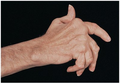
Rheumatoid Arthritis (RA): Chronic, autoimmune disease. Immune system attacks joint tissues, leading to inflammation, swelling, and joint deformity (ankylosis). Treated with anti-inflammatory drugs and immune suppressants.
Gouty Arthritis: Uric acid crystal deposition in joints, especially the big toe. Can lead to joint fusion if untreated. Treated with drugs, hydration, and dietary changes.
Lyme Disease: Bacterial infection from tick bites, can cause joint pain and arthritis. Treated with antibiotics.
Developmental Aspects of Joints
By embryonic week 8, synovial joints resemble adult joints.
Joint size, shape, and flexibility are modified by use.
Aging leads to shortened and weakened ligaments/tendons, increased risk of herniated discs, and higher prevalence of osteoarthritis.
Full-range-of-motion exercise helps postpone joint problems.
