 Back
BackChapter 8: Joints – Structure, Function, and Clinical Aspects
Study Guide - Smart Notes
 Tailored notes based on your materials, expanded with key definitions, examples, and context.
Joints: Classification and Overview
Structural and Functional Classification of Joints
Joints, or articulations, are sites where two or more bones meet. They are classified both structurally and functionally, which determines their mobility and anatomical features.
Structural classification is based on the material binding bones together and the presence or absence of a joint cavity.
Functional classification is based on the amount of movement allowed by the joint.
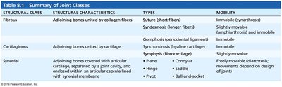
Structural Class | Structural Characteristics | Types | Mobility |
|---|---|---|---|
Fibrous | Adjoining bones united by collagen fibers | Suture (short fiber), Syndesmosis (longer fiber), Gomphosis (periodontal ligament) | Immobile (synarthrosis), Slightly movable, Immobile |
Cartilaginous | Adjoining bones united by cartilage | Synchondrosis (hyaline cartilage), Symphysis (fibrocartilage) | Immobile, Slightly movable |
Synovial | Adjoining bones covered with articular cartilage, separated by a joint cavity, and enclosed within an articular capsule lined with synovial membrane | Plane, Hinge, Pivot, Condylar, Saddle, Ball-and-socket | Freely movable (diarthrosis; movements depend on design of joint) |
Fibrous Joints
Sutures
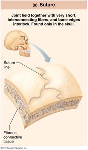
Sutures are rigid, interlocking joints found only in the skull. They are held together by very short, interconnecting fibers and allow for growth during youth. In adults, sutures ossify and become immovable, forming synostoses.
Location: Only in the skull
Movement: Immovable (synarthrosis)
Syndesmoses
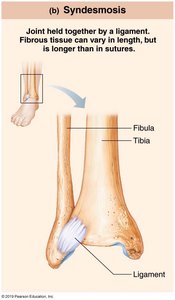
Syndesmoses are joints where bones are connected by ligaments. The length of the fibers determines the degree of movement possible.
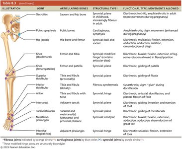
Short fibers: Little to no movement (e.g., inferior tibiofibular joint)
Long fibers: More movement (e.g., interosseous membrane between radius and ulna)
Gomphoses
Gomphoses are peg-in-socket fibrous joints. The only examples are the articulations of the teeth with their sockets, held in place by the periodontal ligament.
Location: Teeth in alveolar sockets
Movement: Immobile
Cartilaginous Joints
Synchondroses
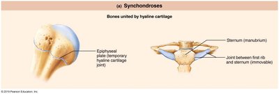
Synchondroses are joints where bones are united by a bar or plate of hyaline cartilage. Most are immovable and serve as temporary joints during development.
Examples: Epiphyseal plates in long bones, joint between first rib and manubrium of sternum
Movement: Immovable (synarthrosis)
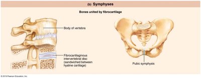
Symphyses
Symphyses are joints where bones are united by fibrocartilage, allowing for strength and flexibility. Hyaline cartilage is also present as articular cartilage on bony surfaces.
Examples: Intervertebral joints, pubic symphysis
Movement: Slightly movable (amphiarthrosis)
Synovial Joints
General Structure of Synovial Joints
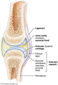
Synovial joints are the most movable type of joint in the body. They are characterized by the presence of a fluid-filled joint cavity and several structural features that allow for a wide range of motion.
Articular cartilage: Hyaline cartilage covering bone ends, preventing crushing of bone ends
Joint (synovial) cavity: Small, fluid-filled space unique to synovial joints
Articular capsule: Two layers—external fibrous layer (dense irregular connective tissue) and inner synovial membrane (produces synovial fluid)
Synovial fluid: Viscous, slippery filtrate that lubricates and nourishes articular cartilage
Reinforcing ligaments: Capsular, extracapsular, and intracapsular ligaments
Nerves and blood vessels: Provide sensory input and nutrients
Movements at Synovial Joints
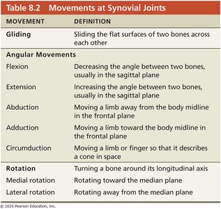
Synovial joints allow for a variety of movements, classified as gliding, angular, or rotational.
Movement | Definition |
|---|---|
Gliding | Sliding the flat surfaces of two bones across each other |
Flexion | Decreasing the angle between two bones, usually in the sagittal plane |
Extension | Increasing the angle between two bones, usually in the sagittal plane |
Abduction | Moving a limb away from the body midline in the frontal plane |
Adduction | Moving a limb toward the body midline in the frontal plane |
Circumduction | Moving a limb or finger so that it describes a cone in space |
Rotation | Turning a bone around its longitudinal axis |
Medial rotation | Rotating toward the median plane |
Lateral rotation | Rotating away from the median plane |
Types of Synovial Joints
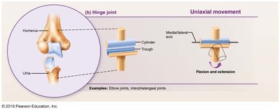
Synovial joints are classified into six types based on the shape of their articular surfaces and the movements they allow:
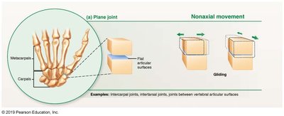
Plane joints: Nonaxial movement (e.g., intercarpal joints)
Hinge joints: Uniaxial movement (e.g., elbow, interphalangeal joints)
Pivot joints: Uniaxial rotation (e.g., proximal radioulnar joint)
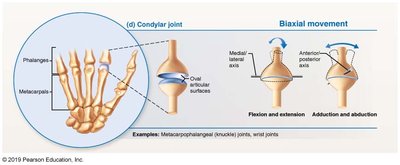
Condylar joints: Biaxial movement (e.g., metacarpophalangeal joints)
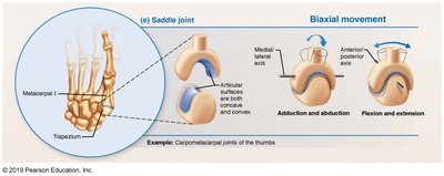
Saddle joints: Biaxial movement (e.g., carpometacarpal joint of thumb)
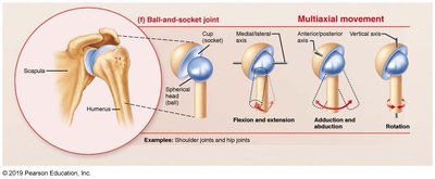
Ball-and-socket joints: Multiaxial movement (e.g., shoulder, hip)
Bursae and Tendon Sheaths
Bursae and tendon sheaths are structures associated with synovial joints that reduce friction and facilitate movement.
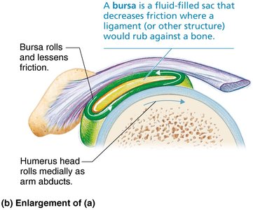
Bursa: A fluid-filled sac that decreases friction where ligaments, muscles, skin, tendons, or bones rub together
Tendon sheath: An elongated bursa that wraps around a tendon subjected to friction
Selected Synovial Joints
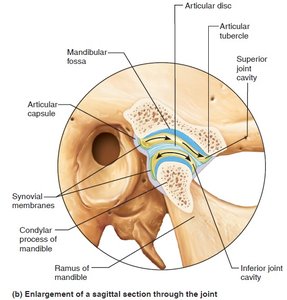
Temporomandibular Joint (TMJ)
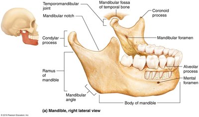
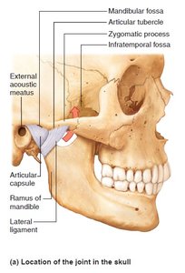
The TMJ is a modified hinge joint that allows both hinge and gliding movements. It is the most easily dislocated joint in the body.
Movements: Depression/elevation (hinge), lateral excursion (gliding)
Articulating bones: Mandibular condyle and temporal bone
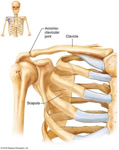
Shoulder (Glenohumeral) Joint
The shoulder joint is a ball-and-socket joint, providing the greatest range of motion in the body. Its stability is sacrificed for mobility.
Articulating bones: Humerus and scapula (glenoid cavity)
Movement: Multiaxial (flexion, extension, abduction, adduction, rotation)
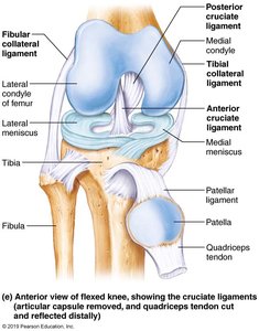
Knee Joint
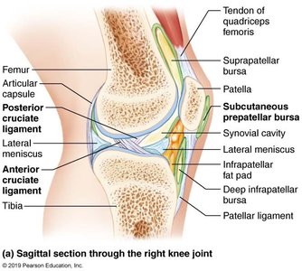
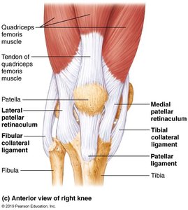
The knee is the largest and most complex joint in the body, consisting of three joints within a single cavity. It allows flexion, extension, and some rotation.
Femoropatellar joint: Plane joint, allows gliding motion
Tibiofemoral joint: Hinge joint, allows flexion, extension, and some rotation
Ligaments: Collateral, cruciate, and patellar ligaments provide stability
Bursae: At least 12 associated with the knee
Common Knee Injuries
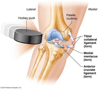
The knee is vulnerable to injury, especially from lateral blows. The "unhappy triad" refers to simultaneous injury of the anterior cruciate ligament (ACL), tibial collateral ligament, and medial meniscus.
Inflammatory and Degenerative Joint Conditions
Bursitis
Inflammation of a bursa, usually caused by trauma or friction. Treated with rest, ice, and anti-inflammatory drugs if severe.
Tendonitis
Inflammation of tendon sheaths, typically due to overuse. Symptoms and treatment are similar to bursitis.
Arthritis
Refers to over 100 types of inflammatory or degenerative diseases that damage joints. Symptoms include pain, stiffness, and swelling. Acute forms are often caused by bacteria and treated with antibiotics; chronic forms include osteoarthritis, rheumatoid arthritis, and gouty arthritis.
Developmental Aspects of Joints
By embryonic week 8, synovial joints resemble adult joints. Joint structure is modified by use, and aging leads to shortening and weakening of ligaments and tendons, with increased risk of joint problems such as osteoarthritis.
Prevention: Full-range-of-motion exercise helps postpone joint problems.
Related Clinical Terms
Arthroplasty: Surgical replacement of a joint
Chondromalacia patellae: Softening and deterioration of the articular cartilage on the posterior surface of the patella
Synovitis: Inflammation of the synovial membrane
