 Back
Chapter 9: Joints – Structured Study Notes
Study Guide - Smart Notes
 Tailored notes based on your materials, expanded with key definitions, examples, and context.
Joints: Classification, Structure, and Function
Section 9-1: Joint Classification and Functional Types
Joints, or articulations, are connections between bones that allow for varying degrees of movement. They are classified by both their structure and their function (range of motion).
Bony Joint Formation: A bony joint (synostosis) forms when two bones fuse and the boundary between them disappears, resulting in an immovable joint. Example: the fusion of frontal bones in the adult skull.
Functional Joint Types:
Synarthrosis: Immovable joint (e.g., sutures of the skull).
Amphiarthrosis: Slightly movable joint (e.g., intervertebral discs).
Diarthrosis: Freely movable joint (e.g., shoulder, knee).
Order of Mobility (Least to Most): Synarthrosis → Amphiarthrosis → Diarthrosis
Structural and Functional Classifications (Table 9–1):
Fibrous Synarthroses:
Suture: Interlocking bones bound by dense connective tissue (e.g., skull sutures).
Gomphosis: Peg-in-socket joint (e.g., teeth in alveolar sockets).
Cartilaginous Synarthrosis: Synchondrosis – Bones united by hyaline cartilage (e.g., epiphyseal plates in children).
Bony Synarthrosis: Synostosis – Bones fused together (e.g., frontal bone).
Fibrous Amphiarthrosis: Syndesmosis – Bones connected by a ligament (e.g., distal tibiofibular joint).
Cartilaginous Amphiarthrosis: Symphysis – Bones joined by fibrocartilage (e.g., pubic symphysis).
Diarthrosis: Synovial joint – Freely movable, with a synovial cavity (e.g., knee, shoulder).
Section 9-2: Synovial Joints (Diarthroses)
Synovial joints are characterized by a joint cavity filled with synovial fluid, surrounded by a joint capsule, and stabilized by accessory structures.
Movement vs. Strength: The more movable a joint, the weaker it is (e.g., the shoulder joint is highly mobile but less stable).
Articular Cartilage: Smooth, slick cartilage covering bone ends reduces friction and absorbs shock.
Synovial Fluid: Viscous fluid within the joint cavity that lubricates, nourishes, and removes waste from articular cartilage.
Nutrient/Waste Exchange: Occurs via diffusion across capillary walls in the areolar tissue of the synovial membrane.
Menisci: Pads of fibrocartilage located in certain joints (e.g., knee) that improve fit and absorb shock.
Fat Pads: Protect articular cartilage and act as packing material.
Sprains: Damage to ligaments stabilizing a joint.
Healing: Bones heal faster than ligaments due to better blood supply.
Bursae: Fluid-filled sacs (with synovial fluid) that reduce friction between tissues.
Bursitis: Inflammation of bursae, often caused by repetitive motion or pressure.
Section 9-3: Types of Synovial Joints and Movements
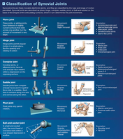
Synovial joints are classified by the shapes of their articulating surfaces and the types of movement they allow.
Plane (Gliding) Joint: Allows slight gliding movements (e.g., intercarpal joints).
Hinge Joint: Allows angular movement in one plane (e.g., elbow, knee).
Condylar Joint: Oval articular face fits within a depression (e.g., radiocarpal joint).
Saddle Joint: Each articular face is concave along one axis and convex along the other (e.g., first carpometacarpal joint).
Pivot Joint: Permits rotation only (e.g., atlantoaxial joint between C1 and C2 vertebrae).
Ball-and-Socket Joint: Allows circumduction and rotation in three planes (e.g., shoulder, hip).
Flexion, Extension, Hyperextension: Flexion decreases the angle between bones; extension increases it; hyperextension extends beyond anatomical position.
Adduction and Abduction: Adduction moves a limb toward the midline; abduction moves it away.
Circumduction: Circular movement combining flexion, extension, abduction, and adduction.
Pronation and Supination: Pronation rotates the forearm so the palm faces down; supination rotates it so the palm faces up. In anatomical position, the forearm is supinated.
Special Movements:
Inversion/Eversion: Inversion turns the sole inward; eversion turns it outward.
Dorsiflexion/Plantar Flexion: Dorsiflexion lifts the foot upward; plantar flexion points the toes downward.
Opposition/Reposition: Opposition moves the thumb to touch other fingers; reposition returns it to anatomical position.
Protraction/Retraction: Protraction moves a body part forward; retraction pulls it back.
Section 9-4: Intervertebral Joints
Intervertebral joints connect adjacent vertebrae and allow for flexibility and movement of the spine.
Types of Joints: Cartilaginous (symphysis) and synovial (facet joints).
Intervertebral Discs: Pads of fibrocartilage between vertebral bodies, composed of an outer anulus fibrosus (fibrocartilage) and an inner nucleus pulposus (gelatinous core for shock absorption).
Height Loss: With age, water loss from discs leads to decreased height.
Ligaments: Strong ligaments stabilize the vertebral column; ligamenta flava connect laminae of adjacent vertebrae.
Section 9-5: Elbow and Knee Joints
The elbow and knee are hinge joints that allow movement in one plane and are stabilized by strong ligaments and menisci.
ACL: Anterior Cruciate Ligament; MCL: Medial Collateral Ligament.
Elbow: The humero-ulnar joint is the largest and strongest articulation, involving the trochlea of the humerus and the trochlear notch of the ulna.
Knee: Medial and lateral menisci are found between femoral and tibial condyles; complete dislocation is rare due to ligament support.
Patella: Embedded in the tendon of the quadriceps femoris.
Ligament Stabilization: The knee is stabilized by the MCL and ACL.
Disc Injuries: A bulging disc is when the disc protrudes but the nucleus pulposus remains contained; a herniated disc is when the nucleus pulposus breaks through the anulus fibrosus.
Section 9-6: Shoulder and Hip Joints
The shoulder and hip are ball-and-socket joints, allowing movement in multiple planes but differing in stability and range of motion.
Shoulder Joint: Glenohumeral joint; most mobile but least stable.
Ligament Stabilization: Acromioclavicular and coracoclavicular ligaments stabilize the shoulder; shoulder separation involves the acromioclavicular joint.
Rotator Cuff Muscles: Supraspinatus, Infraspinatus, Teres Minor, Subscapularis (SITS).
Bursae: Subdeltoid and subcoracoid bursae are found in the shoulder joint.
Hip Joint: Stabilized by iliofemoral and ischiofemoral ligaments; hip fractures can occur at the femoral neck or intertrochanteric region.
Section 9-7: Degenerative Joint Changes
With age, joints may undergo degenerative changes, leading to reduced mobility and pain.
Rheumatism: General term for pain and stiffness in the musculoskeletal system.
Arthritis: Inflammation of joints; most common type is osteoarthritis (degenerative joint disease).
Rheumatoid Arthritis: Autoimmune disorder causing joint inflammation.
Gouty Arthritis: Caused by uric acid crystal deposition in joints.
Section 9-8: Skeletal System Functions
The skeletal system supports the body and stores energy and minerals for other body systems.
