 Back
Chapter 9: Muscles and Muscle Tissue – Comprehensive Study Notes
Study Guide - Smart Notes
 Tailored notes based on your materials, expanded with key definitions, examples, and context.
Muscle Terminology and Types
Muscle Terminology
The scientific study of muscle is known as myology. Muscle fibers are synonymous with muscle cells. Common word roots referring to muscle include myo, mys, and sarco.
Three Types of Muscle Tissue
Muscle tissue is classified into three types based on microscopic anatomy, location, and regulation:
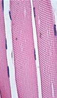
Skeletal Muscle: Voluntary, attached to bones, contracts quickly, fatigues easily.
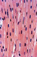
Cardiac Muscle: Involuntary, forms heart wall, autorhythmic, non-fatigable.
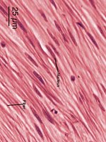
Smooth Muscle: Involuntary, found in hollow organs, contracts slowly, non-fatigable.
Characteristics and Structure of Muscle Tissue
Skeletal Muscle
Skeletal muscle is primarily attached to bones and is under voluntary control. It contracts rapidly and is capable of generating a wide range of forces. The cells are long, cylindrical, striated, and multinucleate.
Cardiac Muscle
Cardiac muscle forms the myocardium of the heart, is involuntary, and exhibits autorhythmicity. The cells are branched, striated, and typically uninucleate, with intercalated discs facilitating rapid excitation.
Smooth Muscle
Smooth muscle is found in the walls of hollow organs and structures. It is involuntary, non-striated, and uninucleate, with slow, sustained contractions.
Functions of Muscle Tissue
Motion: Both external (e.g., walking) and internal (e.g., heartbeat).
Posture: Maintains body posture.
Stabilization: Stabilizes joints.
Thermogenesis: Generates heat through contractions.
Functional Characteristics of Muscle Tissue
Excitability: Ability to respond to stimuli.
Contractility: Ability to shorten.
Extensibility: Ability to stretch without damage.
Elasticity: Ability to return to original shape.
Conductivity: Ability to transmit excitation.
Connective Tissue Wrappings of Skeletal Muscle
Connective Tissue Layers
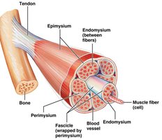
Skeletal muscle is organized by several connective tissue layers:
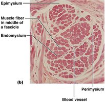
Endomysium: Wraps individual muscle fibers.
Perimysium: Surrounds fascicles (bundles of muscle fibers).
Epimysium: Encloses the entire muscle.
Deep Fascia: Binds groups of muscles.
Superficial Fascia: Located beneath the skin.
Nerve and Blood Supply
Each muscle fiber receives a branch of a motor nerve and is supplied by arteries and veins. Blood vessels branch extensively to provide direct supply to each fiber.
Muscle Attachments
Origin and Insertion
Muscles attach to bones or connective tissue at two points:
Origin: Attached to stationary bone.
Insertion: Attached to moving bone.
Attachment Structure
Direct Attachment: Epimysium fused to periosteum.
Indirect Attachment: Connective tissue forms tendons (cords) or aponeuroses (sheets).
Microscopic Anatomy of Skeletal Muscle Fiber
Muscle Fiber Structure
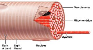
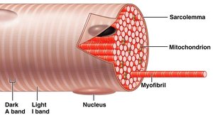
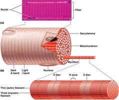
Muscle fibers are long, cylindrical, and multinucleate. The sarcolemma is the cell membrane, and the sarcoplasm is the cytoplasm, rich in myoglobin.
Myofibrils and Sarcomeres
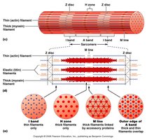
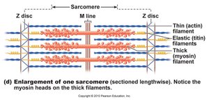
Myofibrils are bundles of contractile protein filaments. The sarcomere is the repeating unit of contraction, defined as the region between two Z discs.
Organelles of Muscle Fiber
Mitochondria: Provide ATP for contraction.
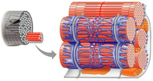
Sarcoplasmic Reticulum (SR): Stores Ca2+ ions.
Myofilaments and Sarcomere Structure
Types of Myofilaments
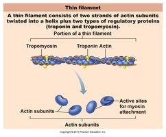
Thin Filaments: Actin, tropomyosin, troponin.
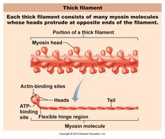
Thick Filaments: Myosin.
Elastic Filaments: Titin, connects myosin to Z discs.
Sarcomere Bands and Lines
The sarcomere contains distinct bands and lines:
Z discs: Boundary between sarcomeres.
A band: Overlap of thick and thin filaments.
I band: Thin filaments only.
H zone: Thick filaments only.
M line: Anchors thick filaments.
Myosin and Actin Proteins
Myosin Protein
Myosin has a rod-like tail and two heads, each with ATPase and actin-binding sites. The heads point to the Z line, tails to the M line. ATP hydrolysis powers the "ratchet" action.
Actin Protein
Actin filaments are helical strands of G actin, each with a myosin-binding site. Tropomyosin spirals around actin, and troponin regulates binding via Ca2+ sites.
Sarcoplasmic Reticulum and T-Tubules
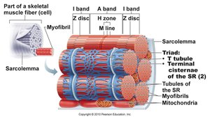
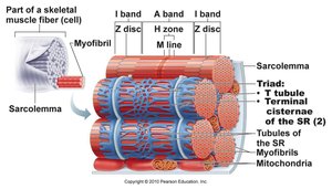
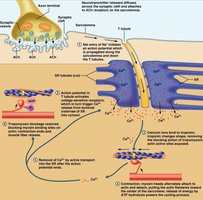
The sarcoplasmic reticulum (SR) stores Ca2+ ions, while T-tubules are inward folds of the sarcolemma that help transmit action potentials.
Muscle Fiber Triads
A triad consists of two terminal cisternae of the SR and one T-tubule, crucial for excitation-contraction coupling.
Neuromuscular Junction and Regulation of Contraction
Neuromuscular Junction Structure
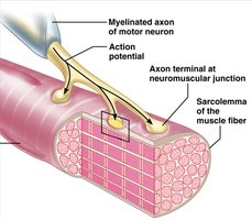
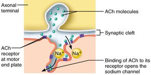
The neuromuscular junction is where motor neurons communicate with muscle fibers, consisting of an axon terminal and motor end plate.
Synaptic Transmission
Acetylcholine (ACh) is released from synaptic vesicles, crosses the synaptic cleft, and binds to ACh receptors, opening Na+ channels and depolarizing the sarcolemma.
Generation of Action Potential
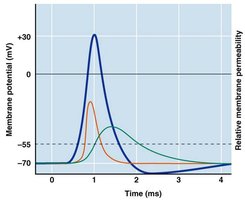
Depolarization opens voltage-gated Na+ channels, triggering an action potential that spreads across the sarcolemma and down T-tubules.
Excitation-Contraction Coupling
The action potential triggers Ca2+ release from the SR, which binds to troponin, causing tropomyosin to move and allowing myosin to bind to actin.
Sliding Filament Model and Cross Bridge Cycling
Sliding Filament Model
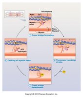
Thin and thick filaments slide past each other, shortening sarcomeres and thus the muscle.
Cross Bridge Cycling
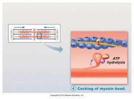
The cycle involves myosin binding to actin, performing a power stroke, releasing actin, and "cocking" the myosin head for the next cycle. ATP hydrolysis provides energy for this process.
Motor Units and Muscle Contraction
Motor Units
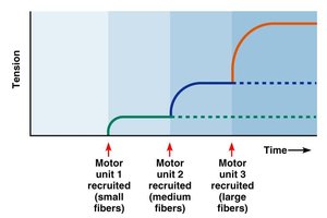
A motor unit consists of a motor neuron and the muscle fibers it innervates. The size of motor units varies, affecting the precision and force of contraction.

Graded Muscle Responses
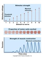
Muscle contraction force can be altered by changing stimulation frequency (temporal summation), stimulus strength (recruitment), and muscle length.
Length-Tension Relationship
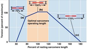
Optimal sarcomere length allows maximum cross bridge formation and tension. Too much or too little overlap reduces tension.
Muscle Tone and Types of Contraction
Isometric Contraction: Tension increases, muscle does not shorten.
Isotonic Contraction: Muscle shortens, tension remains constant.
Muscle Tone: Continuous, passive partial contraction of muscles.
Muscle Metabolism
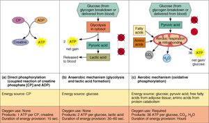
ATP Replenishment Pathways
Creatine Phosphate System: Rapid ATP generation, short duration.
Anaerobic Glycolysis: Produces ATP and lactic acid, short-term.
Aerobic Respiration: Uses O2, long-term ATP supply.
Muscle Fiber Types
Slow Oxidative Fibers: Slow contraction, fatigue resistant, red.
Fast Oxidative Fibers: Fast contraction, moderate fatigue resistance.
Fast Glycolytic Fibers: Fast contraction, easily fatigued, white.
Smooth Muscle Tissue
Structure and Function
Smooth muscle fibers are spindle-shaped, uninucleate, and lack striations. They are organized in longitudinal and circular layers in hollow organs, facilitating peristalsis and segmentation.
Contraction Mechanisms
Smooth muscle contracts under the influence of autonomic nerves, hormones, and local factors. Whole sheets contract in unison due to gap junctions.
Developmental Aspects and Homeostatic Imbalances
Muscle Development
Muscle tissue develops from embryonic mesoderm (myoblasts). Skeletal muscle fibers form by fusion, while cardiac and smooth muscle develop gap junctions.
Regeneration and Aging
Cardiac and skeletal muscle become amitotic but can hypertrophy. Satellite cells provide limited regeneration. Muscle mass decreases with age (sarcopenia), but exercise can reverse this.
Homeostatic Imbalances
Muscular Dystrophy: Inherited diseases causing muscle destruction.
Duchenne Muscular Dystrophy: Lack of dystrophin protein, early onset, severe.
Summary Table: Muscle Types Comparison
Muscle Type | Control | Cell Shape | Striations | Nuclei | Location |
|---|---|---|---|---|---|
Skeletal | Voluntary | Long, cylindrical | Striated | Multinucleate | Bones |
Cardiac | Involuntary | Branched | Striated | Uninucleate | Heart |
Smooth | Involuntary | Spindle-shaped | Non-striated | Uninucleate | Hollow organs |
Additional info: Academic context was added to clarify mechanisms, terminology, and physiological relevance for exam preparation.
