 Back
Chapter 9: Myology – The Muscular System and Skeletal Muscle Tissue
Study Guide - Smart Notes
 Tailored notes based on your materials, expanded with key definitions, examples, and context.
Myology: The Study of Muscle
Introduction to Muscle Tissue
Muscle tissue is specialized for contraction, enabling movement of the body and its parts. There are three main types of muscle tissue, each with distinct structures, locations, and functions.
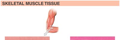
Skeletal Muscle: Voluntary, striated muscle attached to bones and skin. Responsible for movement and heat generation.
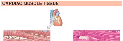
Cardiac Muscle: Involuntary, striated muscle found only in the heart. Features intercalated discs for synchronized contraction.
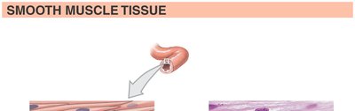
Smooth Muscle: Involuntary, non-striated muscle found in walls of hollow organs and blood vessels. Controls movement of substances and vessel diameter.
Muscle Development
Myogenesis: Formation of Skeletal Muscle
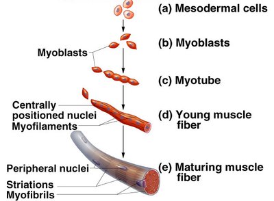
Skeletal muscle fibers develop from embryonic mesodermal cells called myoblasts. These cells fuse to form multinucleated muscle fibers, which mature and organize into functional muscle tissue.
Myoblasts: Embryonic cells that fuse to form muscle fibers.
Myotube: An early, elongated multinucleated cell.
Mature Muscle Fiber: Contains peripheral nuclei, striations, and organized myofibrils.
Levels of Skeletal Muscle Organization
Hierarchical Structure
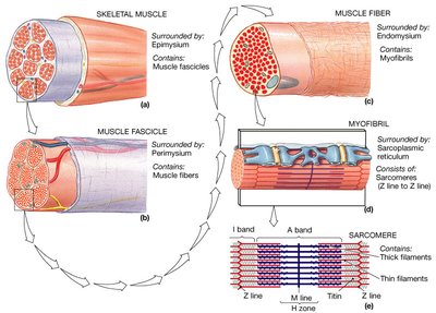
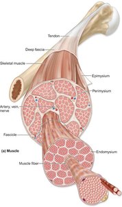
Skeletal muscle is organized into several levels, each surrounded by connective tissue sheaths:
Muscle (organ): Surrounded by epimysium.
Muscle Fascicle: Bundle of muscle fibers, surrounded by perimysium.
Muscle Fiber (cell): Surrounded by endomysium.
Myofibril: Composed of repeating units called sarcomeres.
Sarcomere: The functional contractile unit, made of thick and thin filaments.
Muscle Fiber Anatomy
Internal Structure of a Muscle Fiber
Each muscle fiber contains specialized structures for contraction:
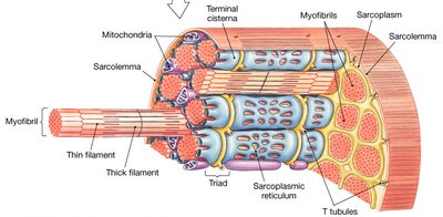
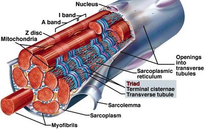
Sarcolemma: The plasma membrane of a muscle fiber.
Sarcoplasm: The cytoplasm, containing organelles and myofibrils.
Myofibrils: Cylindrical structures containing sarcomeres.
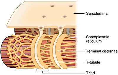
Sarcoplasmic Reticulum (SR): Stores calcium ions; forms terminal cisternae adjacent to T-tubules.
T-tubules (Transverse Tubules): Invaginations of the sarcolemma that transmit action potentials into the fiber.
Triad: A T-tubule flanked by two terminal cisternae of the SR.
Sarcomere Structure and Muscle Proteins
Organization of the Sarcomere
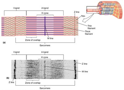
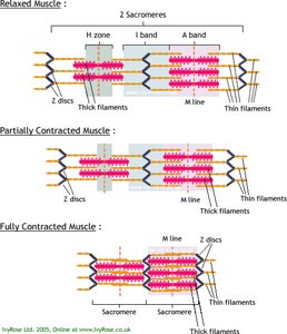
The sarcomere is the basic contractile unit of muscle, defined as the region between two Z lines. It contains overlapping thick and thin filaments, which slide past each other during contraction.
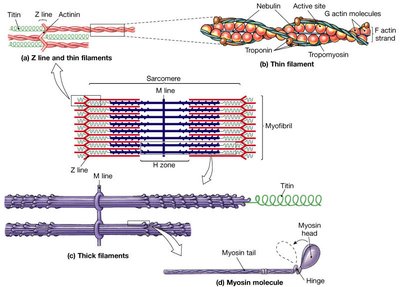
Z line (disc): Anchors thin filaments; marks sarcomere boundaries.
M line: Anchors thick filaments in the center of the sarcomere.
A band: Region containing the entire length of thick filaments.
I band: Region with only thin filaments; spans two adjacent sarcomeres.
H zone: Central region with only thick filaments.
Muscle Filaments and Proteins
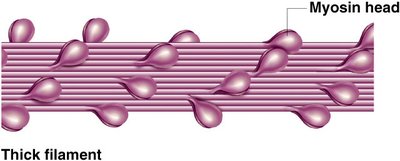
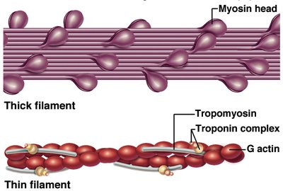
Thick Filaments: Composed mainly of myosin molecules, each with a tail and two heads.
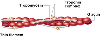
Thin Filaments: Composed of actin, tropomyosin, and troponin complexes.
Contractile Proteins: Myosin (thick) and actin (thin) generate force.
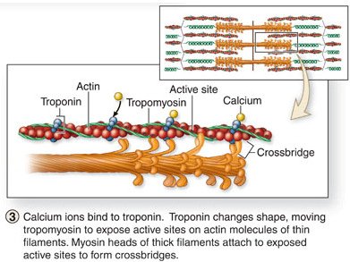
Regulatory Proteins: Tropomyosin blocks binding sites on actin; troponin binds calcium and moves tropomyosin.
Muscle Contraction: Sliding Filament Theory
Mechanism of Contraction
The sliding filament theory explains how muscles contract:
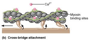
Calcium ions bind to troponin, causing a conformational change that moves tropomyosin and exposes myosin-binding sites on actin.
Myosin heads attach to actin, forming cross-bridges, and pull the thin filaments toward the center of the sarcomere.
The I band and H zone decrease in width, while the A band remains constant. The sarcomere shortens, resulting in muscle contraction.
Key Equations:
Sarcomere length change:
Skeletal Muscle Attachment and Levers
Muscle Attachments
Origin: The fixed attachment point of a muscle.
Insertion: The movable attachment point, pulled toward the origin during contraction.
Belly (body): The thick, central part of the muscle that shortens during contraction.
Lever Systems in the Body
Muscles act on bones as levers to produce movement. The components of a lever system include:
Lever: A rigid bar (bone) that moves on a fixed point (fulcrum).
Fulcrum: The joint around which the lever moves.
Effort: The force applied by muscle contraction.
Resistance (Load): The weight or force that opposes movement.
Lever Class | Arrangement | Example |
|---|---|---|
First Class | Fulcrum in the middle | Neck extension |
Second Class | Resistance in the middle | Standing on tiptoe |
Third Class | Effort in the middle | Biceps curl |
Skeletal Muscle Fiber Organization
Muscle Shapes and Arrangements
The arrangement of muscle fibers affects the muscle's range of motion and power:
Parallel: Fibers run parallel to the long axis (e.g., sartorius).
Convergent: Fibers converge to a single tendon (e.g., pectoralis major).
Circular: Fibers arranged in concentric rings (e.g., orbicularis oris).
Pennate: Fibers attach obliquely to a central tendon.
Unipennate: Fibers on one side of tendon (e.g., extensor digitorum longus).
Bipennate: Fibers on both sides of tendon (e.g., rectus femoris).
Multipennate: Multiple tendons with fibers (e.g., deltoid).
Summary Table: Types of Muscle Tissue
Type | Appearance | Location | Control | Function |
|---|---|---|---|---|
Skeletal | Striated, multinucleated, peripheral nuclei | Attached to bones/skin | Voluntary | Movement, heat |
Cardiac | Striated, branched, single nucleus, intercalated discs | Heart (myocardium) | Involuntary | Pumping blood |
Smooth | Non-striated, single nucleus | Walls of hollow organs, vessels | Involuntary | Movement of substances |
