Back
BackChapter 9: The Muscular System – Structure and Function of Skeletal Muscles
Study Guide - Smart Notes
 Tailored notes based on your materials, expanded with key definitions, examples, and context.
9.1 Structure of Skeletal Muscles
Gross Anatomy of a Skeletal Muscle
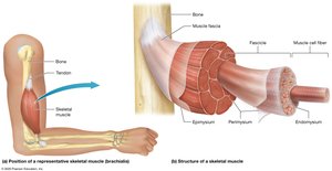
Skeletal muscles are complex organs composed of muscle fibers, connective tissue, blood vessels, and nerves. Understanding their structure is essential for grasping how muscles generate movement and force.
Skeletal Muscle Fibers: Long, thin cells called muscle fibers, surrounded by a thin layer of extracellular matrix known as the endomysium.
Fascicle: A bundle of 10–100 muscle fibers grouped together, surrounded by perimysium (connective tissue).
Epimysium: Connective tissue that surrounds all fascicles in a muscle.
Fascia: The most superficial connective tissue sheath, continuous with the epimysium.
Attachment and Innervation
Tendons: Attach muscle to bone or other structures to facilitate movement.
Blood and Nerve Supply: Skeletal muscles are richly supplied with blood vessels and nerves, essential for function and repair.
Voluntary Control: Skeletal muscle contraction is under conscious control and requires nervous system stimulation.
Muscle Knots (Myofascial Trigger Points)
Myofascial trigger points, or "muscle knots," are localized, irritable spots within the fascia that can cause pain and sustained contraction.
Causes: Repetitive exercise, trauma, stress, disease, or trapped nerves.
Symptoms: Pain and sustained muscle contraction.
Treatment: Massage, anti-inflammatory medications, muscle relaxants, and passive stretching.
Fascicle Patterns and Muscle Shapes
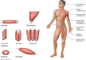
Muscles are classified by the arrangement of their fascicles, which affects their function and appearance.
Parallel: Evenly spaced fascicles, tendon same width as muscle (e.g., Sartorius).
Convergent: Broad at one end, tapers to a single tendon (e.g., Pectoralis major).
Circular (Sphincters): Encircle openings, constrict when contracted (e.g., Orbicularis oculi).
Fusiform: Thick in the middle, tapered at ends (e.g., Biceps brachii).
Pennate: Fascicles attach to tendon at an angle, feather-like appearance:
Unipennate: Fascicles on one side (e.g., Flexor pollicis longus).
Bipennate: Fascicles on both sides (e.g., Rectus femoris).
Multipennate: Several tendons, fascicles join like multiple feathers (e.g., Deltoid).
9.1 Naming Muscles
Principles of Muscle Naming
Muscles are named based on various characteristics to aid in identification and understanding of their function.
Size: Terms like major, minor, longus, brevis, vastus.
Location: Directional (superior, inferior, medial, lateral) and regional anatomical terms (e.g., Pectoralis minor).
Attachment: Named for structures they attach to (e.g., Sternocleidomastoid).
Function: Flexors, extensors, levators, adductors, abductors.
Depth: Profundus (deep), superficialis (superficial).
Common Terms in Muscle Anatomy
Term | Meaning |
|---|---|
Major | Larger |
Minor | Smaller |
Longus | Long |
Brevis | Short |
Vastus | Broad |
Profundus | Deep |
Superficialis | Superficial |
Flexor | Decreases joint angle |
Extensor | Increases joint angle |
Adductor | Moves limb toward midline |
Abductor | Moves limb away from midline |
9.1 Functions of Skeletal Muscles
Muscle Tension and Movement
The primary function of skeletal muscle is to contract and generate muscle tension, which produces movement and heat.
Muscle Tension: Force generated by muscle contraction.
Body Movements: Actions produced by muscle tension.
Heat Generation: Conversion of ATP's chemical energy to mechanical energy produces heat (e.g., shivering).
Functional Groups of Muscles
Muscles work in groups to coordinate movement efficiently and safely.
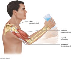
Agonists (Prime Movers): Provide most of the force for a movement.
Antagonists: Oppose and slow the action of agonists, allowing controlled movement.
Synergists: Assist agonists and ensure smooth movement.
Fixators: Stabilize bones, making movement more efficient and reducing injury risk.
Muscle Origin and Insertion
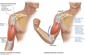
Muscles attach to bones at two points: the origin (more fixed) and the insertion (moves during contraction).
Origin (O): Attachment to the stationary bone.
Insertion (I): Attachment to the bone that moves.
Example: Biceps brachii—origin at scapula, insertion at radius.
Origin and insertion can switch depending on the movement (e.g., iliopsoas during forward bending).
Lever Systems Used in Body Movements
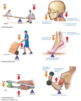
Muscles and bones interact as lever systems to produce movement. There are three classes of levers in the body:
Lever: The bone that moves.
Load: The object or body part being moved.
Force: Muscle tension applied to the lever.
Fulcrum: The joint or hinge point.
First-Class Lever: Fulcrum between load and force (e.g., atlanto-occipital joint).
Second-Class Lever: Load between fulcrum and force (e.g., metatarsophalangeal joints; rare in body).
Third-Class Lever: Force between load and fulcrum (e.g., elbow joint; most common in body).
Mechanical Advantage and Disadvantage
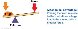
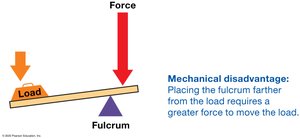
Levers can provide a mechanical advantage (move a large load with less force) or disadvantage (require more force to move a load).
Mechanical Advantage: Fulcrum close to load; small force moves large load.
Mechanical Disadvantage: Fulcrum far from load; greater force needed to move load.
Muscle Strains
A muscle strain occurs when a muscle is overstretched or subjected to excessive force, resulting in tearing of muscle fibers.
Symptoms: Pain, limited movement, swelling, bruising.
Treatment: PRICE method—Protect, Rest, Ice, Compression, Elevation; may include medications, physical therapy, or surgery.

9.1 Studying Muscles
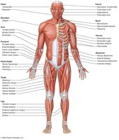
Muscle Groups of the Human Body
Muscles are categorized by their location and function in the body. Understanding these groups aids in learning muscle anatomy.
Muscles of the head, neck, and vertebral column
Muscles of the trunk and pelvic floor
Muscles of the pectoral girdle and upper limb
Muscles of the hip and lower limb
Familiarity with the anterior and posterior superficial muscles provides a foundation for further study.