 Back
BackChromosomal Abnormalities: Number and Structure Changes in Humans
Study Guide - Smart Notes
 Tailored notes based on your materials, expanded with key definitions, examples, and context.
Chromosomal Abnormalities
Introduction
Chromosomal abnormalities are significant alterations in the number or structure of chromosomes, leading to various genetic disorders. These abnormalities can affect physical development, cognitive abilities, and overall health. Understanding these changes is crucial for diagnosing and managing genetic diseases.
Changes in Chromosome Number
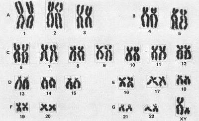
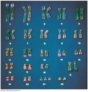
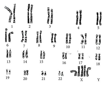
Human Karyotype
A karyotype is an organized profile of a person's chromosomes, arranged and numbered by size from largest to smallest. Humans typically have 46 chromosomes (23 pairs), including one pair of sex chromosomes (XX or XY).
Common Chromosome Number Disorders
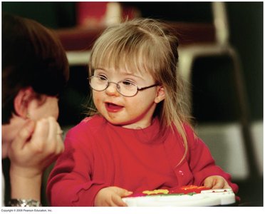
Down Syndrome (Trisomy 21): Caused by an extra copy of chromosome 21. Features include characteristic facial appearance, short stature, heart defects, increased risk of respiratory infections, leukemia, Alzheimer’s disease, and varying degrees of intellectual disability.
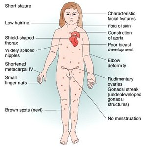
Turner Syndrome (XO): Affects females who have only one X chromosome. Symptoms include short stature, webbed neck, sterility, and normal intelligence.
Klinefelter Syndrome (XXY): Affects males with an extra X chromosome. Features include tall stature, sterility, breast enlargement, and normal intelligence.
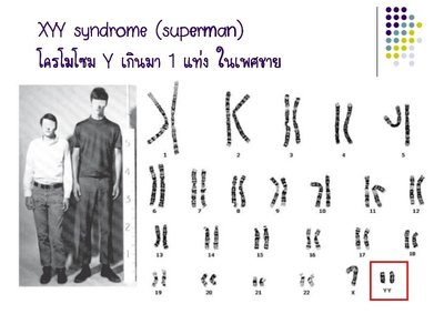
Supermale Syndrome (XYY): Males with an extra Y chromosome. Usually few symptoms, but may include tall stature, acne, and increased risk of learning problems.
Superfemale Syndromes (XXX, XXXX, XXXXX): Females with extra X chromosomes. Symptoms range from mild developmental delays to intellectual disability and craniofacial abnormalities, depending on the number of extra X chromosomes.
Examples of Chromosomal Disorders
Down Syndrome: Most common chromosomal abnormality. Characterized by intellectual disability and distinct physical features.
Turner Syndrome: Features include short stature, webbed neck, and underdeveloped ovaries.
Klinefelter Syndrome: Tall males with breast development and sterility.
XYY Syndrome (Supermale): Tall males, sometimes with learning difficulties.
Superfemale Syndromes: Range from mild to severe intellectual and physical symptoms depending on the number of extra X chromosomes.
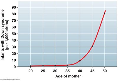
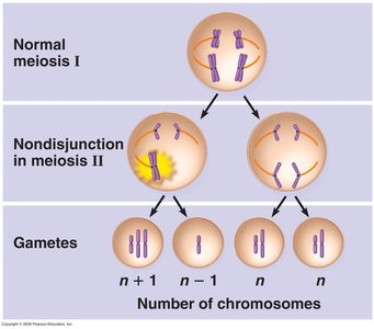
Maternal Age and Nondisjunction
The risk of chromosomal nondisjunction events, such as those leading to Down syndrome, increases with maternal age. Nondisjunction is the failure of chromosomes to separate properly during meiosis, resulting in gametes with abnormal chromosome numbers.
Mechanisms of Chromosome Number Changes
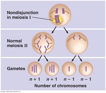
Chromosome number abnormalities often result from nondisjunction during meiosis. This can occur in either meiosis I or II, leading to gametes with extra or missing chromosomes.
Nondisjunction in Meiosis I: Homologous chromosomes fail to separate.
Nondisjunction in Meiosis II: Sister chromatids fail to separate.
Changes in Chromosome Structure
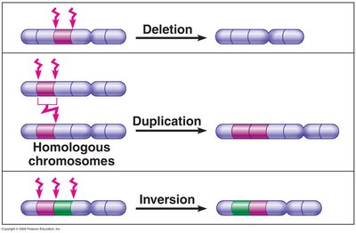
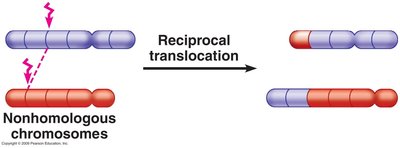
Types of Structural Changes
Structural abnormalities arise from errors during crossing over in meiosis. These include deletions, duplications, inversions, and translocations.
Deletion: Loss of a chromosome segment.
Duplication: Repetition of a chromosome segment.
Inversion: Reversal of a chromosome segment.
Translocation: Movement of a chromosome segment to a nonhomologous chromosome.
Human Disorders Due to Structural Mutations
Translocation Down Syndrome: Part of chromosome 21 is translocated to chromosome 14.
Charcot-Marie-Tooth Disease: Duplication of part of chromosome 17.
Wolf-Hirschhorn Syndrome: Deletion of part of chromosome 4, leading to intellectual disability and physical abnormalities.
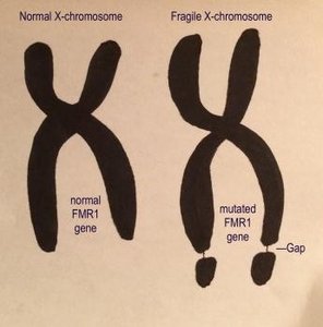
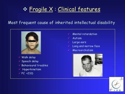
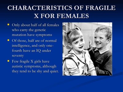
Fragile X Syndrome
Fragile X syndrome (FXS) is the second most common genetic cause of intellectual disability after Down syndrome. It is caused by an expansion of the FMR1 gene on the X chromosome, leading to a lack of the FMRP protein necessary for normal brain development.
Clinical Features: Intellectual disability, autism, large ears, long face, macroorchidism (enlarged testes in males), and behavioral issues.
Genetic Mechanism: Expansion of CGG repeats in the FMR1 gene.
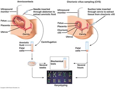
Prenatal Testing for Chromosome Abnormalities
Methods
Prenatal testing can detect chromosomal abnormalities before birth. The two main methods are:
Amniocentesis: Sampling of amniotic fluid to obtain fetal cells for karyotyping.
Chorionic Villus Sampling (CVS): Sampling of placental tissue for earlier diagnosis.
Both procedures carry a small risk of miscarriage.
Summary Table: Common Chromosomal Disorders
Disorder | Karyotype | Main Features |
|---|---|---|
Down Syndrome | 47,XX,+21 or 47,XY,+21 | Intellectual disability, characteristic facial features, heart defects |
Turner Syndrome | 45,X | Short stature, webbed neck, sterility, normal intelligence |
Klinefelter Syndrome | 47,XXY | Tall stature, sterility, breast development, normal intelligence |
XYY Syndrome | 47,XYY | Tall stature, possible learning difficulties |
Triple X Syndrome | 47,XXX | Usually mild symptoms, possible speech/language delays |
Wolf-Hirschhorn Syndrome | del(4p) | Intellectual disability, craniofacial abnormalities |
Fragile X Syndrome | X chromosome with FMR1 expansion | Intellectual disability, autism, behavioral issues |
Additional info: Chromosomal abnormalities can be detected through cytogenetic techniques such as karyotyping, which visually displays the number and structure of chromosomes. Early diagnosis allows for better management and counseling for affected families.
