 Back
Circulation of Blood: Structure, Function, and Pathology of Blood Vessels
Study Guide - Smart Notes
 Tailored notes based on your materials, expanded with key definitions, examples, and context.
Circulation of Blood
Overview of the Circulatory System
The circulatory system is responsible for transporting blood throughout the body, delivering oxygen and nutrients to tissues, and removing waste products. It consists of the heart, blood vessels, and blood. Blood vessels are classified into arteries, veins, and capillaries, each with distinct structures and functions.
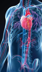
Blood Vessels: Types and Structure
Types of Blood Vessels
Arteries: Carry blood away from the heart toward capillaries. The largest artery is the aorta. Arteries branch into smaller arterioles.
Veins: Carry blood toward the heart from capillaries. Small venules merge to form larger veins, with the superior and inferior vena cava being the largest.
Capillaries: Microscopic vessels that connect arterioles and venules, facilitating exchange of substances between blood and tissues.
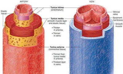
Structure of Blood Vessels
Arteries:
Tunica intima: Inner endothelial layer.
Tunica media: Thick smooth muscle and elastic tissue; regulates blood pressure.
Tunica externa: Outer fibrous connective tissue.
Veins:
Tunica intima: Contains valves to prevent backflow.
Tunica media: Thinner than in arteries.
Tunica externa: Often thicker than in arteries.
Capillaries: Composed of a single layer of endothelial cells (tunica intima) to facilitate exchange.
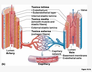
Capillary Beds and Exchange
Capillary beds are networks of capillaries where exchange of gases, nutrients, and wastes occurs between blood and tissues. Precapillary sphincters regulate blood flow into capillary beds.
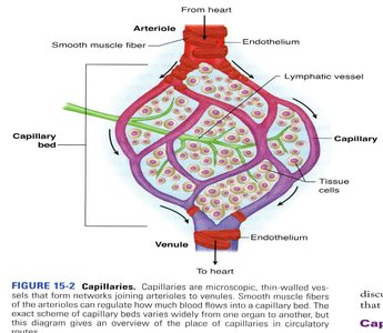
Capillary Exchange Mechanisms
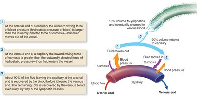
At the arterial end, hydrostatic pressure pushes fluid out of capillaries.
At the venous end, osmotic pressure draws fluid back into capillaries.
About 10% of fluid enters lymphatics and returns to circulation.
Venous Return and Valve Function
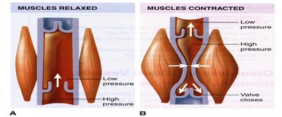
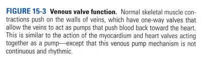
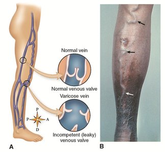
Veins contain one-way valves that prevent backflow. Skeletal muscle contractions help propel blood toward the heart, a mechanism known as the skeletal muscle pump.
Functions of Blood Vessels
Arteries: Distribute oxygen, nutrients, and maintain blood pressure.
Capillaries: Serve as exchange vessels for nutrients, wastes, and fluids.
Veins: Collect blood for return to the heart; operate under low pressure.
Pathological Conditions of Blood Vessels
Arterial Disorders
Arteriosclerosis: Hardening of arteries due to calcification of fatty deposits, reducing blood flow and possibly causing ischemia or necrosis.
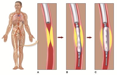
Atherosclerosis: Accumulation of lipids and other matter blocking arteries.
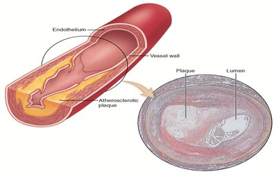
Balloon Angioplasty: A procedure to mechanically widen narrowed arteries.
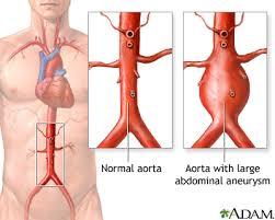
Aneurysm: Abnormal widening of an arterial wall, increasing risk of rupture and hemorrhage.
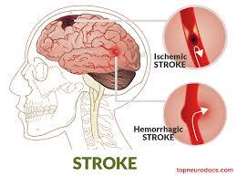
Cerebrovascular Accident (Stroke): Ischemia or hemorrhage in brain tissue due to embolism or vessel rupture.
Venous Disorders
Varicose Veins: Enlarged, twisted veins due to valve failure and blood pooling. Hemorrhoids are varicose veins in the rectum.
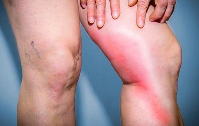
Thrombophlebitis: Inflammation of a vein with clot formation, which can lead to pulmonary embolism.
Routes of Circulation
Systemic and Pulmonary Circulation
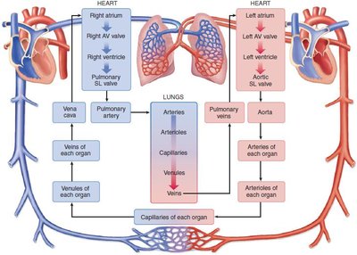
Systemic Circulation: Carries oxygenated blood from the left ventricle through the body and returns deoxygenated blood to the right atrium.
Pulmonary Circulation: Carries deoxygenated blood from the right ventricle to the lungs and returns oxygenated blood to the left atrium.
Hepatic Portal Circulation
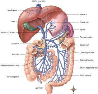
The hepatic portal system is a unique route where blood from the digestive organs passes through the liver before returning to the heart. This allows the liver to process nutrients and detoxify substances.
Fetal Circulation
Fetal circulation includes unique structures such as the placenta, umbilical arteries and vein, ductus venosus, ductus arteriosus, and foramen ovale, which allow the fetus to obtain oxygen and nutrients from maternal blood. At birth, these structures close as the newborn begins to breathe air.
Hemodynamics: Blood Pressure and Flow
Blood Pressure
Definition: The force exerted by blood against vessel walls. Highest in arteries, lowest in veins.
Blood Pressure Gradient: Drives blood flow from high to low pressure regions.
Hypertension (HTN): Blood pressure above 140/90 mm Hg; risk factor for heart disease, kidney failure, and stroke.
Factors Influencing Blood Pressure
Blood Volume: Greater volume increases pressure.
Strength of Heart Contractions: Stronger contractions raise pressure.
Heart Rate: Increased rate raises pressure.
Blood Viscosity: Thicker blood increases pressure.
Peripheral Resistance: Resistance in vessels affects pressure; regulated by vasomotor mechanisms (constriction/dilation).
Venous Return Mechanisms
Strong heart contractions
Adequate arterial pressure
Venous valves
Skeletal muscle pump
Respiratory pump (pressure changes during breathing)
Pulse
The pulse is the rhythmic expansion and recoil of an artery as blood is pumped by the heart. Pulse points are locations where the pulse can be felt, such as the radial, carotid, and femoral arteries.
Circulatory Shock
Circulatory shock is the failure of the circulatory system to deliver adequate oxygen to tissues, leading to cell impairment. Types include:
Cardiogenic Shock: Due to heart failure.
Hypovolemic Shock: Due to blood volume loss (e.g., hemorrhage).
Neurogenic Shock: Due to loss of vascular tone from nerve dysfunction.
Anaphylactic Shock: Due to severe allergic reaction causing vessel dilation.
Septic Shock: Due to toxins from infection causing vessel dilation.
Major Systemic Arteries and Veins
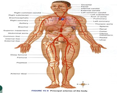
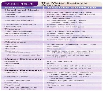
Principal Arteries of the Body
Artery | Tissues Supplied |
|---|---|
Occipital | Posterior head and neck |
Facial | Mouth, pharynx, and face |
Coronary | Heart muscle |
Axillary | Axilla (armpit) |
Femoral | Thigh and leg |
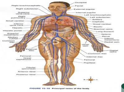
Principal Veins of the Body
Vein | Tissues Drained |
|---|---|
Superior sagittal sinus | Brain |
External jugular | Superficial areas of the head and neck |
Hepatic portal | Abdominal viscera |
Femoral | Leg |
Hepatic Portal Circulation
Summary Table: Comparison of Arteries, Veins, and Capillaries
Feature | Arteries | Veins | Capillaries |
|---|---|---|---|
Direction of Blood Flow | Away from heart | Toward heart | Connect arterioles and venules |
Wall Structure | Thick, muscular, elastic | Thin, less muscular, valves present | Single endothelial layer |
Pressure | High | Low | Very low |
Function | Transport, pressure maintenance | Return, reservoir | Exchange |
