Back
BackClassification and Structure of Tissues: Epithelial and Connective Tissue
Study Guide - Smart Notes
 Tailored notes based on your materials, expanded with key definitions, examples, and context.
Classification of Tissues
Introduction to Tissues
Tissues are groups of cells that share similar structure and function, forming the fundamental building blocks of organs in multicellular organisms. The study of tissues is known as histology. Most complex organisms begin as a single fertilized cell, which divides and differentiates into specialized cells that form tissues.
Cells differentiate into specialized types, such as connective tissue cells and cartilage cells.
Tissues are organized into organs, which perform specific body functions.
Types of Tissues
Epithelial Tissue
Connective Tissue
Muscle Tissue
Nervous Tissue
Epithelial Tissue
Functions and Qualities
Epithelial tissue covers external and internal surfaces of the body, forms glands, and is involved in protection, secretion, absorption, filtration, excretion, and sensory reception.
Cellularity: Composed of closely packed cells with minimal intercellular material.
Avascular: Lacks blood vessels; nutrients are obtained by diffusion from adjacent connective tissue.
Regeneration: Cells can divide rapidly to replace lost or damaged tissue.
Polarity: Has an apical (free) surface and a basal surface attached to a basement membrane.

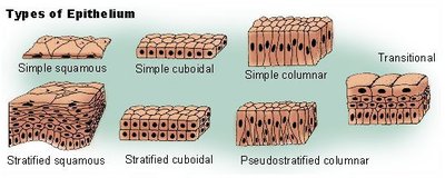
Classification of Epithelial Tissue
By Shape
Squamous: Flat cells
Cuboidal: Cube-shaped cells
Columnar: Tall, column-like cells
By Arrangement
Simple: One cell layer
Stratified: Two or more cell layers
Pseudostratified: Appears stratified but is actually a single layer
Transitional: Stratified epithelium with balloon-shaped cells at the apical surface, capable of stretching
Types of Epithelium
The main types of epithelial tissue are classified by cell shape and arrangement. Each type has distinct functions and locations in the body.
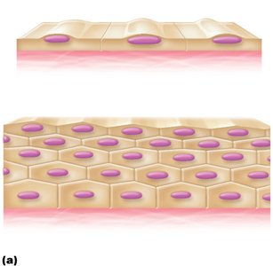
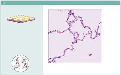
Simple Squamous Epithelium
Description: Single layer of flattened cells with disc-shaped nuclei.
Function: Allows diffusion and filtration; secretes lubricating substances.
Location: Kidney glomeruli, air sacs of lungs, lining of heart, blood vessels, lymphatic vessels, serosae.
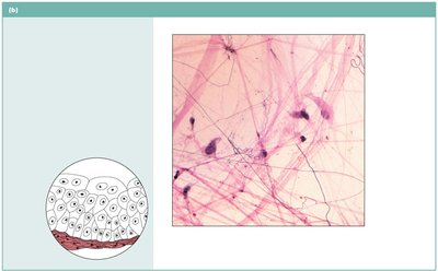
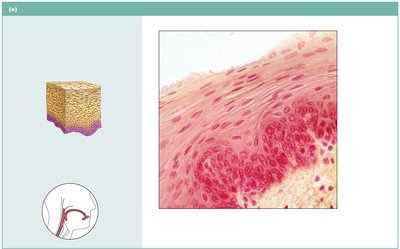
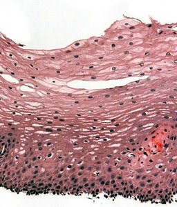
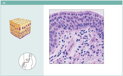
Stratified Squamous Epithelium
Description: Several cell layers; basal cells are cuboidal/columnar, surface cells are flattened.
Function: Protects underlying tissues in areas subjected to abrasion.
Location: Nonkeratinized: esophagus, mouth, vagina; Keratinized: epidermis of skin.
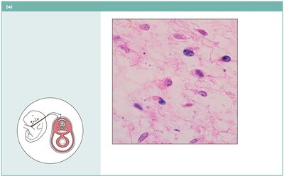
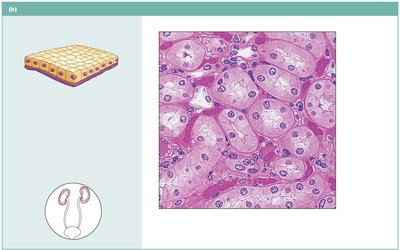
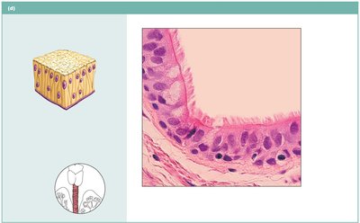
Simple Cuboidal Epithelium
Description: Single layer of cubelike cells with large, spherical nuclei.
Function: Secretion and absorption.
Location: Kidney tubules, ducts and secretory portions of small glands, ovary surface.
Stratified Cuboidal Epithelium
Description: Generally two layers of cubelike cells.
Function: Protection.
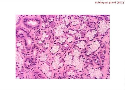
Location: Largest ducts of sweat glands, mammary glands, salivary glands.
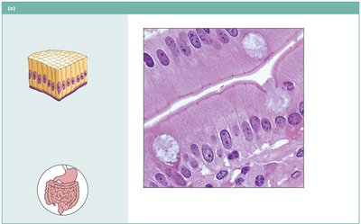
Simple Columnar Epithelium
Description: Single layer of tall cells; may have cilia and goblet cells.
Function: Absorption; secretion of mucus, enzymes; ciliated type propels mucus.
Location: Nonciliated: digestive tract, gallbladder; Ciliated: small bronchi, uterine tubes.
Stratified Columnar Epithelium
Description: Several cell layers; superficial cells elongated and columnar.
Function: Protection; secretion.
Location: Rare; male urethra, large ducts of some glands.
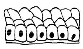
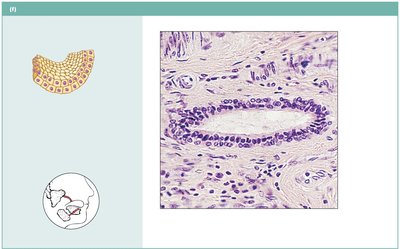
Pseudostratified Columnar Epithelium
Description: Single layer of cells of differing heights; nuclei at different levels; may have cilia and goblet cells.
Function: Secretes mucus; propulsion of mucus by ciliary action.
Location: Nonciliated: sperm-carrying ducts; Ciliated: trachea, upper respiratory tract.
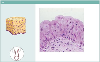
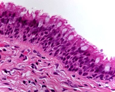
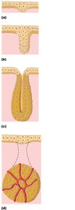
Transitional Epithelium
Description: Resembles both stratified squamous and cuboidal; surface cells dome-shaped.
Function: Stretches readily and permits distension of urinary organs.
Location: Ureters, urinary bladder, part of urethra.
Glandular Epithelium
Epithelial cells forming glands are specialized to manufacture and secrete materials. Glands are classified as exocrine or endocrine based on their method of secretion.
Exocrine glands: Deliver products to an epithelial surface via a duct.
Endocrine glands: Ductless; produce hormones released into extracellular fluid and then into the bloodstream or lymphatic system.
Exocrine Glands
Serous glands: Secrete thin, watery fluid; stain darkly.
Mucous glands: Secrete thick, slippery mucus; stain lightly.
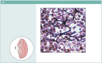
Connective Tissue
Functions and Characteristics
Connective tissue is the most abundant tissue type, providing support, filling spaces, connecting tissues, protecting against infection, and aiding in tissue repair. It is composed of relatively few cells and an extensive extracellular matrix.
Provides support and framework
Fills spaces and connects tissues
Protects against infection
Helps repair tissue damage
Structure of Connective Tissue
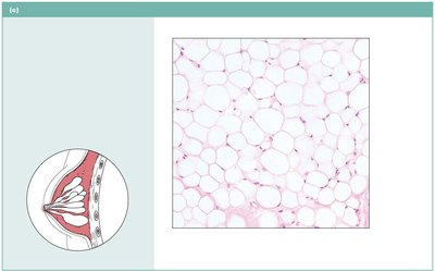
Cells: Most common is the fibroblast; also includes adipocytes, chondrocytes, osteocytes, mast cells, and macrophages.
Extracellular Matrix: Composed of fibers (collagen, elastic, reticular) and ground substance (fluid, gel, or solid filled with proteoglycans and glycoproteins).
Classification of Connective Tissue
Embryonic Connective Tissue: Mesenchyme gives rise to other connective tissue types.
Connective Tissue Proper: Loose (areolar, adipose, reticular) and dense (regular, irregular, elastic).
Supporting Connective Tissue: Cartilage and bone.
Fluid Connective Tissue: Blood and lymph.