 Back
BackCollege Study Guide: Tissues, Integumentary System, and Bone Structure
Study Guide - Smart Notes
 Tailored notes based on your materials, expanded with key definitions, examples, and context.
Chapter 4: Tissue Level of Organization
Epithelial Tissues
Epithelial tissues cover body surfaces, line cavities, and form glands. They are classified by cell shape and number of layers, and are specialized for protection, absorption, secretion, and sensation.
Key Features: Cellularity, polarity, attachment to basement membrane, avascularity, and high regeneration rate.
Specializations: Cilia, microvilli, secretory cells, and a germinative (basal) layer.
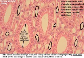
Simple Squamous Epithelium
Structure: Single layer of flat cells.
Locations: Mesothelia, endothelia, alveoli, kidney tubules, cornea.
Functions: Diffusion, filtration, reduces friction, controls vessel permeability.
Simple Cuboidal Epithelium
Structure: Single layer of cube-shaped cells.
Locations: Glands, ducts, kidney tubules, thyroid gland.
Functions: Secretion, absorption, limited protection.
Simple Columnar Epithelium
Structure: Tall, column-shaped cells, often with microvilli or goblet cells.
Locations: Stomach, intestine, gallbladder, uterine tubes, collecting ducts.
Functions: Protection, secretion, absorption.
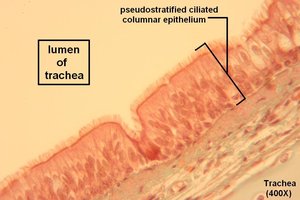
Pseudostratified Columnar Epithelium
Structure: Appears layered but all cells touch the basement membrane; often ciliated with goblet cells.
Locations: Nasal cavity, trachea, bronchi, male reproductive tract.
Functions: Protection, secretion, movement of mucus by cilia.
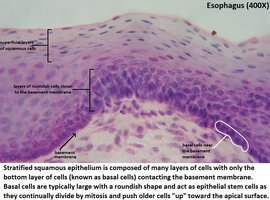
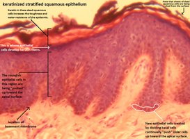
Stratified Squamous Epithelium
Structure: Multiple layers; surface cells are flat.
Locations: Skin (keratinized), mouth, throat, esophagus, rectum, anus, vagina (non-keratinized).
Functions: Physical protection against abrasion, pathogens, and chemical attack.
Keratinized: Adds strength and water resistance (skin).
Non-keratinized: Stays moist (oral cavity, esophagus, vagina).
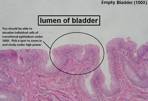
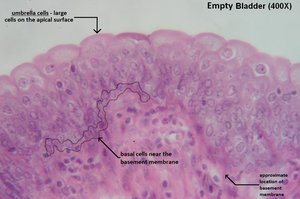
Transitional Epithelium
Structure: Multiple layers; cells change shape (dome-shaped when relaxed, flattened when stretched).
Locations: Urinary bladder, renal pelvis, ureters.
Function: Permits expansion and recoil after stretching.
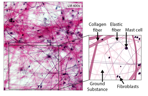
Connective Tissues (CT)
Connective tissues provide support, store energy, and transport materials. They are characterized by specialized cells and an extracellular matrix (fibers + ground substance).
Cell Types: Fibroblasts, adipocytes, macrophages.
Fiber Types: Collagen (strength), elastic (stretch), reticular (network).
Areolar (Loose) Connective Tissue
Structure: Gel-like ground substance, loosely organized fibers, many blood vessels.
Locations: Under epithelia, around organs.
Functions: Connects, protects, stores water/salts/glucose.
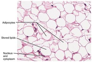
Adipose Tissue
Structure: Large, closely packed adipocytes (fat cells) with nucleus pushed to the side.
Locations: Deep to skin, around eyes/kidneys.
Functions: Padding, insulation, energy storage.
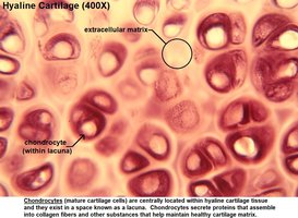
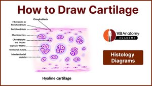
Supportive Connective Tissue: Cartilage
General Function: Shock absorption and protection.
Structure: Chondrocytes in lacunae, avascular, surrounded by perichondrium.
Hyaline Cartilage
Locations: Rib-sternum, ends of bones at joints, larynx, trachea, bronchi, nasal septum.
Functions: Stiff but flexible support, reduces friction between bones.
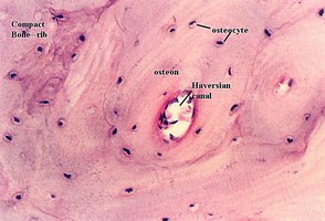
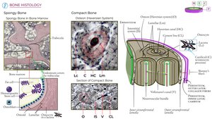
Supportive Connective Tissue: Bone
Compact Bone: Dense, organized in osteons (Haversian systems); strong and weight-bearing.
Spongy Bone: Trabeculae with marrow spaces; lighter and supports marrow.
Bone Cells: Osteocytes in lacunae, connected by canaliculi.
Chapter 5: Integumentary System (Skin)
Overview and Functions
The integumentary system includes the skin and its accessory structures (hair, nails, glands). It protects the body, regulates temperature, synthesizes vitamin D, and provides sensory information.
Functions: Protection, excretion, storage, sensation, vitamin D synthesis, dehydration prevention, temperature regulation.
Skin Layers
Epidermis: Keratinized stratified squamous epithelium; main cell = keratinocyte.
Dermis: Connective tissue (papillary = areolar CT; reticular = dense irregular CT).
Hypodermis: Adipose + areolar tissue; binds skin to muscle, insulation.

Epidermal Layers (Deep to Superficial)
Stratum basale/germinativum: Mitotic stem cells; bonds to dermis.
Stratum spinosum: Living keratinocytes; Langerhans cells.
Stratum granulosum: Cells produce keratin; stop dividing.
Stratum lucidum: Only in thick skin; translucent.
Stratum corneum: Dead, flat, keratinized cells; desquamation (shedding).
Other Epidermal Cells
Melanocytes: Produce melanin for UV protection.
Dendritic (Langerhans) cells: Immune defense.
Tactile (Merkel) cells: Sensation of touch.
Skin Color Factors
Hemoglobin (blood), carotene (diet), melanin (genetics/UV exposure).
Clinical indicators: Cyanosis (blue, low O2), erythema (red, inflammation), jaundice (yellow, liver).
Accessory Structures
Hair: Protection, insulation, sensation.
Sebaceous glands: Secrete sebum (oil).
Sudoriferous glands: Eccrine (watery sweat), apocrine (thicker, odor).
Nails: Keratinized cells for protection and manipulation.
Burns and Skin Cancer
Burns: 1st degree (epidermis), 2nd degree (epidermis + dermis), 3rd degree (full thickness).
Skin Cancer: Basal cell carcinoma, squamous cell carcinoma, malignant melanoma (ABCDE rule).
Chapter 6: Osseous Tissue & Bone Structure
Functions of Bone
Support, protection, movement, storage (minerals/fat), hematopoiesis (blood cell formation).
Classification and Structure
Shapes: Long, short, flat, irregular, sesamoid, sutural.
Long Bone Structure: Diaphysis (shaft), epiphysis (ends), metaphysis (growth zone).
Osseous Tissue Composition
Matrix: 2/3 inorganic (hydroxyapatite), 1/3 organic (collagen).
Cells: Osteoprogenitor, osteoblasts, osteocytes, osteoclasts.
Bone Development & Growth
Osteogenesis: Bone formation.
Intramembranous ossification: Flat bones (skull).
Endochondral ossification: Most bones; cartilage model replaced by bone.
Epiphyseal plate: Growth in length; closes to form epiphyseal line in adults.
Bone Homeostasis
Balance between osteoblast (build) and osteoclast (breakdown) activity.
Regulated by exercise, nutrition (Ca2+, phosphate, vitamins C, D, A), and hormones (PTH, calcitonin, calcitriol).
Calcium Balance
Critical for nerve and muscle function.
Regulated by parathyroid hormone (raises Ca2+), calcitonin (lowers Ca2+), and calcitriol.
Aging and Bone Disorders
Osteopenia: Bone thinning with age.
Osteoporosis: Severe bone loss, increased fracture risk.
Quick Exam Tips
Memorize tissue locations and functions.
Know skin layers and color factors.
Understand bone matrix and cell types.
Practice identifying tissues from images.
