Back
BackCommunication, Integration, and Homeostasis: Study Notes for ANP College Students
Study Guide - Smart Notes
 Tailored notes based on your materials, expanded with key definitions, examples, and context.
Communication, Integration, and Homeostasis
Overview of Cell-to-Cell Communication
Cell-to-cell communication is essential for coordinating physiological functions and maintaining homeostasis. Cells use both chemical and electrical signals to interact locally and over long distances.
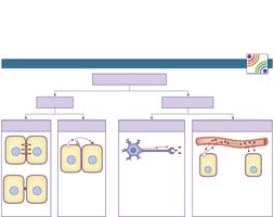
Local Communication: Occurs between adjacent or nearby cells using gap junctions, contact-dependent signals, autocrine, and paracrine signaling.
Long-Distance Communication: Utilizes electrical signals (neurons) and chemical signals (hormones, cytokines, neurohormones, extracellular vesicles) that travel through the circulatory system.
Local Communication Mechanisms
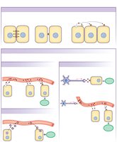
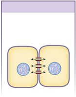
Gap Junctions: Direct cytoplasmic connections between adjacent cells allow ions and small molecules to pass freely, enabling rapid communication.
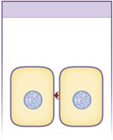
Contact-Dependent Signals: Require interaction between membrane-bound molecules on two cells.
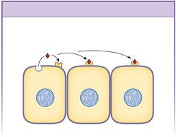
Autocrine Signals: Act on the same cell that secreted them.
Paracrine Signals: Secreted by one cell and diffuse to adjacent cells.
Long-Distance Communication Mechanisms
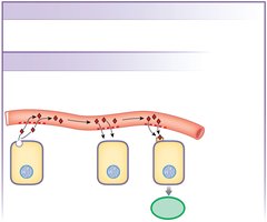
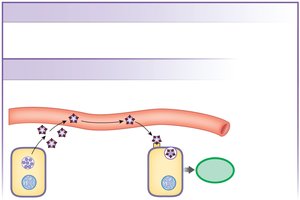
Endocrine System: Hormones and cytokines are secreted into the blood and travel throughout the body. Only target cells with specific receptors respond.
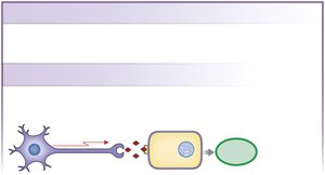
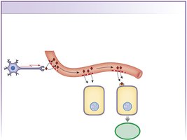
Nervous System: Neurotransmitters are released by neurons and diffuse across synapses to target cells. Neurohormones are released into the blood for action at distant targets.
Extracellular Vesicles: Released by cells to carry signals to distant targets.
Communication Among Systems
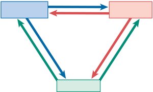
The endocrine, nervous, and immune systems interact through shared signaling molecules such as hormones, neurocrine molecules, and cytokines.
Signal Pathways and Receptors
General Steps in Signal Pathways
Most signal pathways follow a sequence of steps:
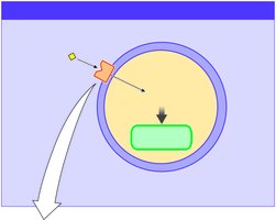
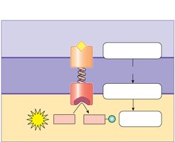
Signal molecule binds to a membrane receptor protein.
Receptor activates intracellular signal molecules.
Intracellular signals alter target proteins.
Target proteins create a cellular response.
Types of Target Cell Receptors
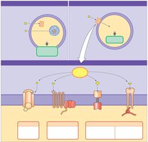
Intracellular Signal Receptors: Lipophilic (fat-soluble) signal molecules diffuse through the cell membrane and bind to cytosolic or nuclear receptors, triggering slower responses related to gene activity.
Cell Membrane Receptors: Extracellular (water-soluble) signal molecules bind to membrane receptors, triggering rapid cellular responses.
Categories of Membrane Receptors:
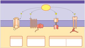
Receptor-channel: Ligand binding opens or closes ion channels.
G protein–coupled receptor (GPCR): Ligand binding opens ion channels or alters enzyme activity.
Receptor-enzyme: Ligand binding activates an intracellular enzyme.
Integrin receptor: Ligand binding alters enzymes or the cytoskeleton.
Signal Transduction and Amplification
Signal Transduction
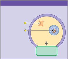
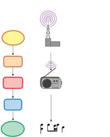
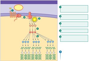
Signal transduction converts one form of signal into another, often amplifying the signal and creating a cascade of intracellular events.
Transducer: Converts the external signal into an intracellular response.
Amplifier: Increases the strength of the signal.
Biological Signal Transduction Pathways
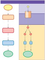
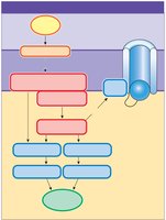
Signal molecule binds to membrane receptor protein.
Receptor activates intracellular signal molecules (second messengers).
Second messengers alter target proteins, leading to a cellular response.
Amplifier enzymes and protein kinases play key roles in these pathways.
Signal Cascades and Amplification
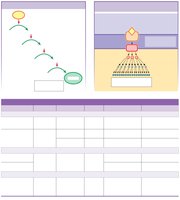
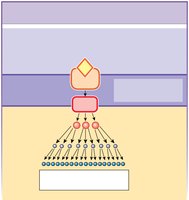
Cascade: Sequential activation of proteins, where each step activates the next.
Amplification: A single ligand can result in the production of many intracellular molecules, greatly increasing the effect.
Second Messenger Pathways
Second messengers are intracellular molecules that relay signals from receptors to target proteins. Common second messengers include cAMP, cGMP, IP3, DAG, and Ca2+.
Second Messenger | Made From | Amplifier Enzyme | Linked To | Action | Effects |
|---|---|---|---|---|---|
cAMP | ATP | Adenylyl cyclase | GPCR | Activates protein kinases, binds ion channels | Phosphorylates proteins, alters channel opening |
cGMP | GTP | Guanylyl cyclase | Receptor-enzyme | Activates protein kinases | Phosphorylates proteins |
IP3 | Membrane phospholipids | Phospholipase C | GPCR | Releases Ca2+ from stores | Ca2+ effects |
DAG | Membrane phospholipids | Phospholipase C | GPCR | Activates protein kinase C | Phosphorylates proteins |
Ca2+ | Ca2+ stores | Various | Various | Binds to calmodulin, other proteins | Alters enzyme activity, exocytosis, contraction |

Signal Transduction Using Ion Channels
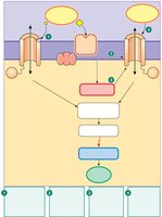
Receptor-channels open or close in response to signal molecule binding.
Some channels are directly linked to G proteins; others respond to second messengers or electrical/mechanical signals.
Changes in ion permeability create electrical signals and cellular responses.
G Protein-Coupled Signal Transduction
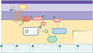
GPCR-Adenylyl Cyclase Pathway: Signal molecule binds to GPCR, activating G protein, which turns on adenylyl cyclase. Adenylyl cyclase converts ATP to cAMP, which activates protein kinase A, leading to phosphorylation of proteins and a cellular response.
GPCR-Phospholipase C Pathway: Signal molecule activates GPCR and G protein, which activates phospholipase C. PLC converts membrane phospholipids into DAG and IP3. DAG activates protein kinase C; IP3 releases Ca2+ from organelles, creating a Ca2+ signal.
Receptor-Enzyme Signaling: Tyrosine Kinase
Tyrosine kinase transfers a phosphate group from ATP to a tyrosine residue of a protein, activating the protein.
Signal molecule binds to surface receptor, activating tyrosine kinase on the cytoplasmic side.
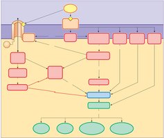
Summary Map of Signal Transduction
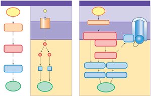
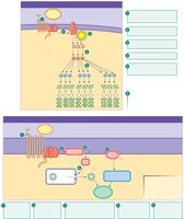
Signal transduction involves multiple steps, including changes in ion concentration, activation of G proteins, amplifier enzymes, protein kinases, and altered proteins, leading to various cellular responses.
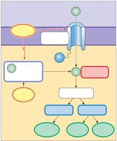
Calcium as an Intracellular Messenger
Ca2+ enters the cell through voltage-gated channels or is released from intracellular stores.
Ca2+ binds to proteins such as calmodulin, altering protein activity and triggering processes like exocytosis and muscle contraction.
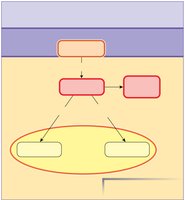
Arachidonic Acid Cascade
Membrane phospholipids are converted to arachidonic acid by phospholipase A2.
Arachidonic acid is further processed by lipoxygenase and cyclooxygenase (COX) to produce lipid-soluble paracrines such as leukotrienes, prostaglandins, and thromboxanes.
Receptor Agonists and Antagonists
Agonist: Activates the receptor, mimicking the primary ligand.
Antagonist: Blocks receptor activity, preventing a response.
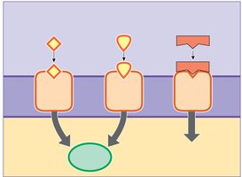
Target Response Depends on Receptor Type
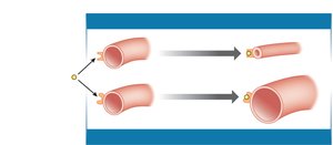
The same ligand (e.g., epinephrine) can produce different responses depending on the receptor isoform present on the target cell.
Example: Epinephrine causes constriction in intestinal blood vessels (α-receptor) and dilation in skeletal muscle blood vessels (β2-receptor).
Control Patterns in Physiology
Tonic and Antagonistic Control
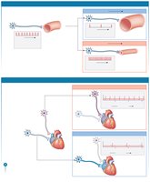
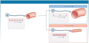
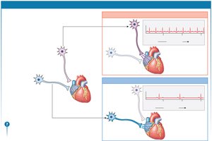
Tonic Control: Regulates physiological parameters in an up-down fashion; the signal is always present but changes in intensity.
Antagonistic Control: Uses different signals to send a parameter in opposite directions (e.g., sympathetic and parasympathetic neurons controlling heart rate).
Reflex Pathways and Homeostasis
Steps in a Reflex Pathway
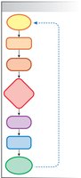
Long-distance pathways use response loops to maintain homeostasis. The steps include:
Stimulus
Sensor
Input signal
Integrating center
Output signal
Target
Response
Feedback loop
Multiple Meanings of the Word Receptor
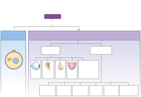
Receptor: Can refer to a protein that binds a ligand or a specialized cell/structure for transduction of stimuli into electrical signals.
Sensory receptors are classified as central (in or near the brain) or peripheral (outside the brain).
Simple and Complex Reflexes
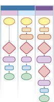
Simple Reflexes: Have one integrating center (neural or endocrine).
Complex Reflexes: Involve two or more integrating centers (neuro-endocrine).
Comparison of Neural and Endocrine Control
Neural and endocrine reflexes differ in their pathways, integrating centers, and output signals. Complex neuroendocrine reflexes involve multiple integrating centers and hormones.
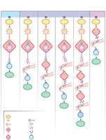
Chapter Summary
Chemical signals can be amplified.
Receptors may have multiple ligands; one ligand may have multiple receptors.
Cells can up-regulate or down-regulate their receptors to enhance or depress their response.
Signal pathways have mechanisms for termination.
Tonic control allows pathways to change intensity.
Communication occurs locally and over long distances, using direct contact, diffusion, neurons, and blood.
Signal molecules (first messengers) interact with intracellular or cell membrane receptors.
Intracellular receptors respond to lipophilic signals, altering gene activity.
Cell membrane receptors respond to lipophobic signals, triggering second messenger systems and rapid responses.
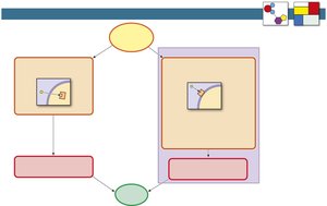
Reflex pathways involve stimuli, sensors, integrating centers, output signals, targets, and responses.
Responses can be described at cellular, tissue, organ, or systemic levels.