 Back
BackCompartmentation: Cells and Tissues – Structured Study Notes
Study Guide - Smart Notes
 Tailored notes based on your materials, expanded with key definitions, examples, and context.
Compartmentation: Cells and Tissues
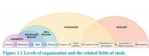
Introduction to Physiology and Cellular Organization
Physiology is the study of normal organismal functioning and its component parts. The cell is the basic functional unit of living organisms, capable of carrying out all processes of life, but no single cell can perform all functions of the mature human body. Cells assemble into tissues, which work together to achieve common purposes.
Cell: Basic unit of life; performs essential functions.
Tissue: Group of cells working together for a specific function.
Organ: Structure composed of multiple tissue types.
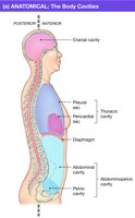
Functional Compartments of the Body
The human body is organized into three major cavities, each surrounded by specific tissue layers and serving distinct functions.
Cranial cavity: Enclosed by bone; contains the brain.
Thoracic cavity: Surrounded by ribs and diaphragm; contains heart and lungs.
Abdominopelvic cavity: Surrounded by peritoneum; contains digestive and reproductive organs.
Fluid-Filled Body Compartments
Body compartments also include fluid-filled spaces such as the circulatory system, eyes, cerebrospinal fluid, and pleural/pericardial sacs. These compartments are essential for physiological processes and protection of organs.
Lumens of Hollow Organs
The lumen is the interior space of any hollow organ, such as the heart, lungs, blood vessels, and intestines. Lumens may be filled with air or fluid and, in some cases, are extensions of the external environment (e.g., the digestive tract).
Lumen: Interior of hollow organs; can be filled with air or fluid.
Example: The lumen of blood vessels is filled with blood.
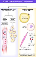
The Body’s Fluid Compartments
Body fluids are divided into intracellular fluid (ICF) and extracellular fluid (ECF). The ECF is further subdivided into plasma and interstitial fluid. These compartments are separated by cell membranes.
Intracellular fluid (ICF): Fluid inside cells.
Extracellular fluid (ECF): Fluid outside cells; includes plasma and interstitial fluid.
Cell membrane: Separates ICF from ECF.
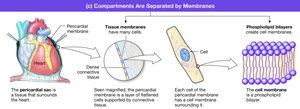
Cell Membranes
Structure and Function of Cell Membranes
Cell membranes are thin layers of lipids that separate the aqueous environments inside and outside the cell. They consist of a phospholipid bilayer and are also known as plasma membranes or plasmalemma.
Physical isolation: Separates ECF from ICF.
Regulation of exchange: Controls entry/exit of ions, nutrients, and waste.
Communication: Contains proteins for environmental response.
Structural support: Anchors cell to cytoskeleton and extracellular matrix.
Membrane Composition
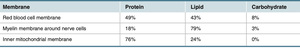
Membranes are composed mainly of lipids and proteins, with the ratio varying by cell type. Metabolically active cells have higher protein content.
Membrane | Protein | Lipid | Carbohydrate |
|---|---|---|---|
Red blood cell membrane | 49% | 43% | 8% |
Myelin membrane around nerve cells | 18% | 79% | 3% |
Inner mitochondrial membrane | 76% | 24% | 0% |
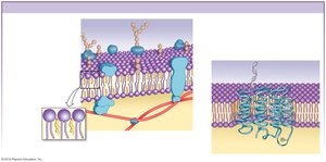
Fluid Mosaic Model
The fluid mosaic model describes the cell membrane as a dynamic structure composed of phospholipids, proteins, glycoproteins, glycolipids, and cholesterol. These components are not fixed and can move within the membrane.
Phospholipids: Form the bilayer.
Proteins: Integral and peripheral; serve various functions.
Carbohydrates: Attach to lipids/proteins; form glycocalyx.
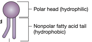
Membrane Lipids and Hydrophobic Barrier
Phospholipids create a hydrophobic barrier that prevents water-soluble (hydrophilic) molecules from crossing without assistance.
Hydrophilic head: Faces water.
Hydrophobic tail: Faces inward, away from water.
Membrane Proteins
Membrane proteins are classified as integral (tightly bound) or peripheral (loosely bound). Integral proteins include transmembrane and lipid-anchored proteins, while peripheral proteins attach via noncovalent interactions.
Integral proteins: Span the membrane or are anchored to lipids.
Peripheral proteins: Attach to membrane proteins; include enzymes and structural proteins.
Membrane Carbohydrates
Carbohydrates are found on the external surface of the cell membrane, attached to proteins (glycoproteins) or lipids (glycolipids). They form the glycocalyx, which is important for immune response and cell recognition.
Glycocalyx: Protective layer formed by membrane carbohydrates.
Example: ABO blood groups are determined by membrane sugars.
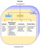
Intracellular Compartments
Cell Structure and Components
Cells are divided into compartments: cell membrane, nucleus, and cytoplasm. The cytoplasm contains cytosol, inclusions, protein fibers (cytoskeleton), and organelles.
Cytosol: Fluid portion of cytoplasm.
Inclusions: Insoluble materials (e.g., ribosomes, nutrients).
Protein fibers: Cytoskeleton for structural support.
Organelles: Membrane-bound structures with specialized functions.
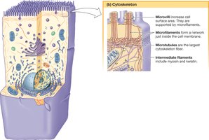
Cytoplasmic Protein Fibers
The cytoskeleton is composed of three types of protein fibers, classified by diameter and protein composition.
Diameter | Type of Protein | Functions |
|---|---|---|
7 nm | Actin (microfilaments) | Muscle contraction, cytoskeleton |
10 nm | Keratin (intermediate filaments) | Hair, nails, protective barrier |
25 nm | Tubulin (microtubules) | Movement of cilia, flagella, chromosomes; intracellular transport |
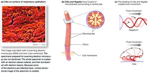
Microtubules, Cilia, and Flagella
Microtubules form cilia and flagella, which facilitate cellular motion. Both structures have a 9+2 arrangement and use the motor protein dynein.
Cilia: Beat rhythmically; move fluids in airways and reproductive tract.
Flagella: Undulate; sperm is the only human cell with flagellum.
Cytoskeleton Functions
The cytoskeleton is a dynamic scaffold that determines cell shape, internal organization, intracellular transport, assembly of cells into tissues, and movement.
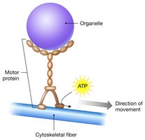
Motor Proteins
Motor proteins use ATP to move along cytoskeletal fibers. Three groups are myosins (muscle contraction), kinesins and dyneins (vesicle movement), and dyneins (cilia and flagella movement).
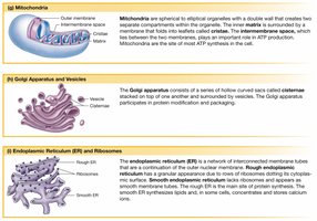
Organelles and Specialized Functions
Organelles are membrane-bound compartments that perform specialized functions. Key organelles include mitochondria (ATP production), endoplasmic reticulum (synthesis, storage, transport), Golgi apparatus (protein modification and packaging), and lysosomes (intracellular digestion).
Mitochondria: Site of cellular respiration and ATP synthesis.
Endoplasmic reticulum: RER synthesizes proteins; SER synthesizes lipids.
Golgi apparatus: Modifies and packages proteins.
Lysosomes: Digestive enzymes; autophagy and phagocytosis.
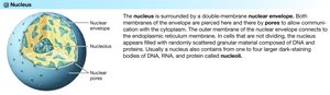
Nucleus
The nucleus is the cell's control center, containing chromatin and directing all cellular functions. The nuclear envelope is a double membrane with pores for communication with the cytoplasm. The nucleolus is the site of ribosomal RNA synthesis.
Tissues of the Body
Histology and Tissue Types
Histology is the study of tissue structure and function. The body has four primary tissue types: epithelial, connective, muscle, and neural.
Epithelial: Protection and regulation of material exchange.
Connective: Structural support and defense.
Muscle: Contraction and movement.
Neural: Electrical signaling and information transfer.
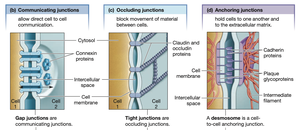
Cell Junctions
Cells in tissues are held together by three types of junctions: communicating (gap), occluding (tight), and anchoring junctions.
Gap junctions: Direct cell-to-cell communication.
Tight junctions: Block movement of materials between cells.
Anchoring junctions: Hold cells to each other and the extracellular matrix.
Epithelial Tissue
Epithelial tissue forms layers around surfaces and can be simple (one cell thick) or stratified (multiple layers). The apical membrane faces the lumen, while the basolateral membrane faces the extracellular fluid. All substances entering or leaving the body must pass through epithelial tissue.
Functional Categories of Epithelia
Exchange epithelium: Thin, flattened cells for gas exchange; simple squamous epithelium.
Transporting epithelium: Cuboidal/columnar cells; specialized for transport.
Ciliated epithelium: Cilia move fluids/particles in respiratory and reproductive tracts.
Protective epithelium: Prevents exchange; protects against stress.
Secretory epithelium: Produces and releases substances; includes exocrine and endocrine glands.
Connective, Muscle, and Neural Tissues
Connective tissue: Includes blood, support tissues, cartilage, and bone; provides structural support and defense.
Muscle tissue: Contracts to produce force and movement; includes cardiac, smooth, and skeletal muscle.
Neural tissue: Neurons transmit signals; glial cells support neurons.
Organs and Integration of Tissues
Organs
Organs are structures composed of multiple tissue types working together. The skin is an example of an organ integrating epithelial, connective, muscle, and neural tissues.
Apoptosis
Programmed Cell Death
Apoptosis is the process of programmed cell death, where cells shrink and break into membrane-bound blebs that are removed by neighboring or immune cells. This process is essential for tissue homeostasis and development.
