 Back
BackComprehensive Study Guide: Central Nervous System, Cardiovascular System, and Endocrine System
Study Guide - Smart Notes
 Tailored notes based on your materials, expanded with key definitions, examples, and context.
Central Nervous System
Brain Regions and Ventricles
The brain is divided into several major regions, each with distinct functions. The ventricles are interconnected cavities within the brain filled with cerebrospinal fluid (CSF), which cushions and protects neural tissue.
Lateral Ventricles: Paired structures located in each cerebral hemisphere.
Interventricular Foramen: Connects lateral ventricles to the third ventricle.
Third Ventricle: Located in the midline, surrounded by the diencephalon.
Cerebral Aqueduct: Narrow channel connecting the third and fourth ventricles.
Fourth Ventricle: Located between the brainstem and cerebellum.
Lateral and Median Apertures: Openings that allow CSF to flow into the subarachnoid space.
Subarachnoid Space: Area between arachnoid mater and pia mater, filled with CSF.
Cerebral Cortex Functional Areas
The cerebral cortex is organized into three functional areas:
Motor Areas: Control voluntary movements.
Sensory Areas: Receive and interpret sensory information.
Association Areas: Integrate information for complex functions.
Cerebral White Matter and Basal Nuclei
Cerebral White Matter: Composed of myelinated fibers that facilitate communication between brain regions.
Basal Nuclei: Groups of neurons involved in regulating voluntary motor movements and procedural learning.
Diencephalon and Brain Stem
Thalamus: Relay station for sensory information.
Hypothalamus: Regulates homeostasis, endocrine functions, and autonomic control.
Epithalamus (Pineal Gland): Secretes melatonin, regulates circadian rhythms.
Midbrain, Pons, Medulla Oblongata: Control vital functions such as respiration, heart rate, and reflexes.
Meninges and Cerebrospinal Fluid
The meninges are protective membranes surrounding the brain and spinal cord.
Dura Mater: Tough, outermost layer.
Arachnoid Mater: Middle layer, contains villi for CSF absorption.
Pia Mater: Delicate, innermost layer.
Cerebrospinal Fluid: Produced by choroid plexus, circulates in ventricles and subarachnoid space, provides protection and nutrient transport.
Cardiovascular System: The Heart
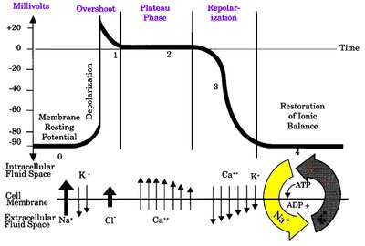
Cardiac Muscle Action Potential
Cardiac muscle cells exhibit a unique action potential characterized by rapid depolarization, a plateau phase, and repolarization. This ensures coordinated contraction and relaxation.
Depolarization: Rapid influx of Na+ ions.
Overshoot: Membrane potential briefly becomes positive.
Plateau Phase: Ca2+ influx maintains depolarization.
Repolarization: K+ efflux restores resting potential.
Restoration of Ionic Balance: Na+/K+ ATPase pump reestablishes ion gradients.
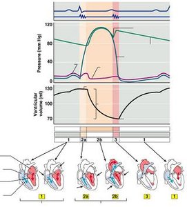
Cardiac Cycle and Heart Function
The cardiac cycle consists of systole (contraction) and diastole (relaxation), regulating blood flow through the heart chambers and valves.
Isovolumetric Contraction: Ventricles contract with all valves closed.
Ventricular Ejection: Semilunar valves open, blood is pumped out.
Isovolumetric Relaxation: Ventricles relax, all valves closed.
Ventricular Filling: AV valves open, blood flows into ventricles.
Electrical Conduction System of the Heart
The heart's electrical system coordinates contraction via specialized pacemaker cells and conduction pathways.
Sinoatrial (SA) Node: Primary pacemaker, initiates impulse.
Atrioventricular (AV) Node: Delays impulse, allows atrial contraction.
AV Bundle (Bundle of His): Conducts impulse to ventricles.
Right and Left Bundle Branches: Carry impulse through interventricular septum.
Purkinje Fibers: Distribute impulse to ventricular myocardium.
Cardiac Output and Stroke Volume
Cardiac Output (CO): Volume of blood pumped per minute.
Stroke Volume (SV): Volume of blood ejected per beat.
Preload: Degree of stretch of cardiac muscle before contraction (Frank-Starling Law).
Afterload: Resistance the heart must overcome to eject blood.
Endocrine System
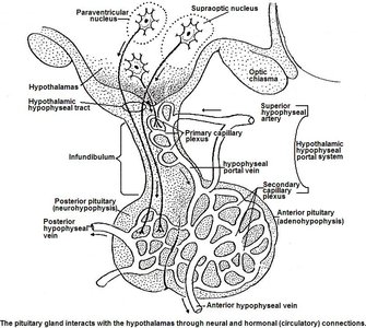
Hypothalamus and Pituitary Gland
The hypothalamus and pituitary gland form a major neuroendocrine control center, regulating hormone release throughout the body.
Hypothalamus: Produces releasing and inhibiting hormones.
Anterior Pituitary (Adenohypophysis): Receives signals via the hypophyseal portal system.
Posterior Pituitary (Neurohypophysis): Stores and releases hormones produced by hypothalamic neurons.
Hypothalamic-Hypophyseal Tract: Neural pathway connecting hypothalamus to posterior pituitary.
Hypophyseal Portal System: Vascular pathway connecting hypothalamus to anterior pituitary.
Hormone Classification and Action
Hormones: Chemical messengers secreted by endocrine glands.
Classification: Based on chemical structure: amino acid-based, steroid, and peptide hormones.
Target Cell Activation: Depends on receptor presence, hormone concentration, and affinity.
Second Messenger System: Protein hormones act via cAMP pathway.
Direct Gene Activation: Steroid hormones bind intracellular receptors, modulate gene expression.
Hypothalamic-Pituitary Hormones
Anterior Pituitary: GH, TSH, ACTH, Prolactin, FSH, LH
Posterior Pituitary: ADH, Oxytocin
Regulation: Hormonal, humoral, and neural stimuli control hormone release.
Clinical Correlations
Hormone Imbalances: Dwarfism, gigantism, acromegaly, diabetes insipidus.
Thyroid Disorders: Cretinism, myxedema, Grave's disease, goiter.
Adrenal Disorders: Addison's disease, Cushing's syndrome, adrenogenital syndrome.
Diabetes Mellitus: Type I and II, complications include hyperglycemia, dehydration, and metabolic acidosis.
Summary Table: Major Pituitary Hormones
Hormone | Target Cells | Function |
|---|---|---|
Growth Hormone (GH) | Body tissues | Stimulates growth and metabolism |
Thyroid Stimulating Hormone (TSH) | Thyroid gland | Stimulates thyroid hormone release |
Adrenocorticotropic Hormone (ACTH) | Adrenal cortex | Stimulates cortisol release |
Prolactin | Mammary glands | Promotes milk production |
Follicle Stimulating Hormone (FSH) | Ovaries/Testes | Stimulates gamete production |
Luteinizing Hormone (LH) | Ovaries/Testes | Stimulates hormone production |
Antidiuretic Hormone (ADH) | Kidneys | Promotes water retention |
Oxytocin | Uterus/Mammary glands | Stimulates contraction and milk ejection |
Additional info: Academic context was added to expand brief points and ensure completeness. Images included are directly relevant to the explanation of cardiac muscle action potential, cardiac cycle, and hypothalamic-pituitary connections.
