 Back
BackComprehensive Study Guide for Anatomy and Physiology: Cardiovascular, Respiratory, Digestive, Metabolic, Renal, and Reproductive Systems
Study Guide - Smart Notes
 Tailored notes based on your materials, expanded with key definitions, examples, and context.
Cardiovascular System
Blood Components and Functions
The cardiovascular system is responsible for transporting blood, nutrients, gases, and waste products throughout the body. Blood consists of plasma, red blood cells (RBCs), white blood cells (WBCs), and platelets.
Plasma: The liquid portion of blood containing water, proteins, electrolytes, and waste products.
RBCs: Carry oxygen via hemoglobin; produced through erythropoiesis.
WBCs: Defend against infection; include neutrophils, lymphocytes, eosinophils, etc.
Platelets: Essential for blood clotting.
Pathophysiology: Anemia, Sickle Cell Disease, DVT, PE, Myocardial Infarction
Anemia: Reduced RBCs or hemoglobin; causes fatigue, pallor, and weakness. Types include iron-deficiency, sickle cell, and hemolytic anemia.
Sickle Cell Disease: Genetic disorder causing abnormal hemoglobin; leads to pain crises and organ damage.
Deep Venous Thrombosis (DVT): Formation of blood clots in deep veins, often in the legs.
Pulmonary Embolism (PE): Clot travels to lungs, causing respiratory distress.
Myocardial Infarction: Heart attack due to blocked coronary artery.
Coagulation and Hemostasis
Hemostasis: Process of stopping bleeding, involving vasoconstriction, platelet plug formation, and clotting cascade.
Clotting Cascade: Series of enzymatic reactions leading to fibrin formation.
Platelet Plug: Platelets adhere to damaged vessel and aggregate.
The Heart: Structure and Function
Pericardium: Protective sac around the heart.
Epicardium, Myocardium, Endocardium: Layers of the heart wall.
Stroke Volume: Amount of blood pumped per beat.
Frank-Starling Law: Increased preload increases stroke volume.
Electrical System of the Heart
Pacemaker Cells: Initiate electrical impulses.
Contractile Cells: Respond to impulses and contract.
EKG: Measures electrical activity; includes P wave, QRS complex, T wave.
Blood Typing
Antigens: Surface markers on RBCs.
Antibodies: Proteins that recognize antigens.
Agglutination: Clumping of RBCs when incompatible blood is mixed.
Respiratory System
Pulmonary Anatomy and Physiology
The respiratory system enables gas exchange and consists of the airways, lungs, and associated structures.
Primary, Secondary, Tertiary Bronchi: Branching airways leading to alveoli.
Alveoli: Site of gas exchange.
Type 1 Pneumocytes: Thin cells for gas exchange.
Type 2 Pneumocytes: Produce surfactant to reduce surface tension.
Goblet Cells: Produce mucus.
Cilia: Move mucus and debris out of airways.
Gas Exchange and Regulation
External Respiration: Gas exchange in lungs.
Internal Respiration: Gas exchange in tissues.
Hemoglobin Saturation: Percentage of hemoglobin bound to oxygen.
Oxygen-Hemoglobin Dissociation Curve: Shows relationship between oxygen partial pressure and hemoglobin saturation.
Bohr Effect: Increased CO2 lowers hemoglobin's affinity for O2.
Haldane Effect: Deoxygenated hemoglobin binds CO2 more readily.
Chloride Shift: Exchange of bicarbonate and chloride ions in RBCs.
Digestive System
Anatomy of the Digestive Tract
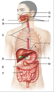
The digestive system breaks down food, absorbs nutrients, and eliminates waste. It consists of the mouth, esophagus, stomach, intestines, and accessory organs.
Route of Food: Mouth → Pharynx → Esophagus → Stomach → Small Intestine (duodenum, jejunum, ileum) → Large Intestine (ascending, transverse, descending colon, rectum) → Anus.
Small Intestine: Duodenum, Jejunum, Ileum
The small intestine is divided into three regions, each with specialized functions in digestion and absorption.
Duodenum: First segment; receives chyme and digestive enzymes.
Jejunum: Middle segment; primary site for nutrient absorption.
Ileum: Final segment; absorbs remaining nutrients and bile salts.
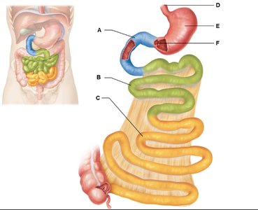
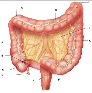
Large Intestine: Colon, Rectum, Mesentery
The large intestine absorbs water and electrolytes, forms feces, and includes the ascending, transverse, descending colon, rectum, and mesentery.
Ascending Colon: First section; absorbs water.
Transverse Colon: Middle section; continues absorption.
Descending Colon: Final section; stores feces.
Rectum: Terminal segment; stores feces before elimination.
Mesentery: Connective tissue anchoring intestines.
Digestive Enzymes and Functions
Parietal Cells: Produce stomach acid (HCl).
Chief Cells: Secrete pepsinogen.
Cholecystokinin (CCK): Stimulates bile and pancreatic enzyme release.
Somatostatin: Inhibits gastric secretion.
Serotonin: Regulates gut motility.
Salivary Amylase: Begins carbohydrate digestion in mouth.
Pancreatic Amylase: Continues carbohydrate digestion in small intestine.
Lactase: Breaks down lactose; deficiency causes lactose intolerance.
Pepsin: Digests proteins in stomach.
Lipase: Digests fats.
Bile: Emulsifies fats.
Liver: Produces bile, processes nutrients.
Gallbladder: Stores and releases bile.
Pancreas: Produces digestive enzymes and juices.
Enteric Nervous System: Controls gut motility.
Peristalsis: Wave-like muscle contractions moving food.
Digestion and Absorption Pathways
Carbohydrate Digestion: Mouth (salivary amylase) → Stomach → Small intestine (pancreatic amylase, brush border enzymes) → Absorbed into bloodstream.
Protein Digestion: Stomach (pepsin) → Small intestine (pancreatic proteases, brush border enzymes) → Absorbed into bloodstream.
Fat Digestion: Small intestine (bile, pancreatic lipase) → Absorbed into lymphatic vessels.
Metabolism and Nutrition
Cellular Respiration
Aerobic Respiration: Requires oxygen; produces more ATP.
Anaerobic Respiration: Occurs without oxygen; produces less ATP.
ATP: Main energy currency of the cell.
NAD/NADH and FAD/FADH2: Electron carriers in cellular respiration.
Glycolysis: Converts glucose to pyruvate; produces ATP and NADH.
Krebs Cycle: Oxidizes acetyl CoA; produces NADH, FADH2, CO2.
Electron Transport Chain: Generates ATP via oxidative phosphorylation.
Proton Gradient: Drives ATP synthesis.
Role of Oxygen: Final electron acceptor in ETC.
Fed and Fasted States
Insulin: Produced by pancreas; promotes glucose uptake and storage.
Glucagon: Produced by pancreas; stimulates glucose release.
Hepatocyte: Liver cell.
Adipocyte: Fat cell.
Glycogen: Storage form of glucose.
Glycogenolysis: Breakdown of glycogen.
Proteolysis: Breakdown of proteins.
Lipolysis: Breakdown of fats.
Gluconeogenesis: Creation of new glucose from non-carbohydrate sources.
Renal System and Homeostasis
Kidney Function and Regulation
Filtration: Removal of waste from blood.
Reabsorption: Return of useful substances to blood.
Secretion: Addition of waste to filtrate.
Excretion: Elimination of waste as urine.
RAAS System: Regulates blood pressure and fluid balance.
Aldosterone, ADH, ANP: Hormones controlling kidney function.
GFR Regulation: Controlled by arteriole dilation/constriction, hormones, and nervous system.
Reproductive System
Anatomy and Physiology
Male and Female Reproductive Systems: Structures and functions for reproduction.
Pregnancy: Physiological changes affecting multiple systems.
Hormonal Regulation: Controls reproductive function and GFR.
Immunity
Innate vs Acquired Immunity
Innate Immunity: Non-specific, immediate defense.
Acquired Immunity: Specific, develops after exposure.
Antibodies and Immune Cells
IgG: Main antibody; crosses placenta.
IgM: First responder.
IgA: Mucosal immunity; in breast milk.
IgE: Allergies and parasites.
Neutrophils: Increased in bacterial infections.
Lymphocytes: Increased in viral infections.
Eosinophils: Increased in parasitic and allergic reactions.
Primary vs Secondary Immune Response
Primary Response: First exposure; slower, less antibody production.
Secondary Response: Subsequent exposure; faster, more robust antibody production.
Additional Info
Spelling is critical for anatomical terms.
Review all structures in provided figures for lab quizzes and exams.
