 Back
Comprehensive Study Guide: Human Anatomy & Physiology Laboratory Workbook (BIO 112)
Study Guide - Smart Notes
 Tailored notes based on your materials, expanded with key definitions, examples, and context.
The Human Body: An Orientation
Anatomical Position and Its Importance
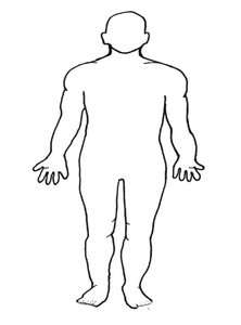
The anatomical position is a standardized posture used in anatomy to ensure consistency when describing locations and directions on the human body. In this position, the body stands upright, facing forward, with arms at the sides and palms facing forward. Feet are flat and slightly apart.
Key Point 1: Anatomical position provides a reference for directional terms and anatomical descriptions.
Key Point 2: It eliminates confusion by standardizing the orientation of the body.
Example: When describing the location of the heart, it is always referenced relative to the anatomical position.
Directional and Regional Terms
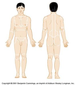
Directional terms are used to describe the location of one body part relative to another. Regional terms specify distinct areas of the body.
Superior: Toward the head
Inferior: Away from the head
Anterior (Ventral): Toward the front/belly side
Posterior (Dorsal): Toward the back
Medial: Toward the midline
Lateral: Away from the midline
Proximal: Closer to the point of attachment
Distal: Farther from the point of attachment
Superficial: Toward the surface
Deep: Away from the surface
Common regional terms include: brachial (arm), gluteal (buttocks), femoral (thigh), patellar (knee), orbital (eye), umbilical (navel).
Body Planes and Sections
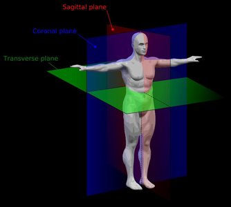
Body planes are imaginary lines used to divide the body for anatomical study.
Sagittal Plane: Divides the body into left and right portions.
Mid-sagittal Plane: Divides the body into equal left and right halves.
Transverse Plane: Divides the body into superior and inferior portions.
Coronal (Frontal) Plane: Divides the body into anterior and posterior portions.
Body Cavities
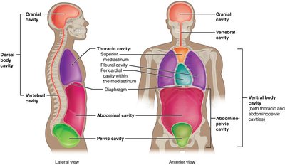
The human body contains several cavities that house organs and provide protection.
Ventral Body Cavity: Located on the anterior side; includes the thoracic and abdominopelvic cavities.
Thoracic Cavity: Contains heart (pericardial cavity), lungs (pleural cavities), and mediastinum.
Abdominopelvic Cavity: Contains abdominal (digestive organs) and pelvic (bladder, reproductive organs) cavities.
Dorsal Body Cavity: Located on the posterior side; includes cranial (brain) and vertebral (spinal cord) cavities.
Cells and Tissues
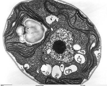
Cellular Organelles and Functions
Cells are the basic units of life, and organelles are specialized structures within cells that perform distinct functions.
Plasma Membrane: Controls passage of materials.
Cytoplasm: Contains organelles and solutes.
Nucleus: Contains DNA; controls cell activities.
Rough Endoplasmic Reticulum (RER): Protein synthesis.
Smooth Endoplasmic Reticulum (SER): Membrane production.
Golgi Apparatus: Modifies and packages proteins.
Mitochondria: Aerobic respiration.
Centrioles: Spindle formation during mitosis.
The Cell Cycle and Mitosis
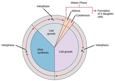
The cell cycle consists of interphase (cell growth and DNA replication) and mitosis (cell division). Mitosis includes prophase, metaphase, anaphase, and telophase, followed by cytokinesis.
Interphase: Cell grows and replicates DNA.
Prophase: Chromatin condenses, nucleus breaks apart.
Metaphase: Chromosomes align at cell equator.
Anaphase: Sister chromatids separate.
Telophase: Nucleus reforms, chromosomes decondense.
Cytokinesis: Division of cytoplasm.
Skin and Body Membranes
Histology and Tissue Types
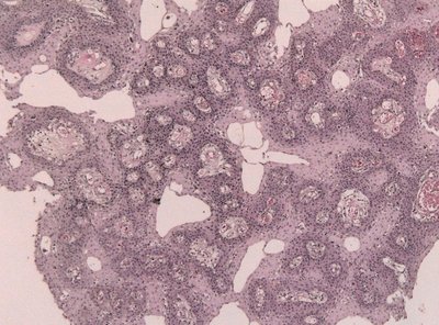
Histology is the study of tissues. The body contains four main tissue types: epithelial, connective, muscle, and nervous.
Epithelial Tissue: Covers surfaces; types include simple squamous, cuboidal, columnar, and stratified squamous.
Connective Tissue: Supports and binds; types include loose areolar, adipose, dense regular/irregular, cartilage, bone, blood.
Muscle Tissue: Movement; types include skeletal, cardiac, and smooth.
Nervous Tissue: Communication; found in brain and spinal cord.
Skin Layers and Structures
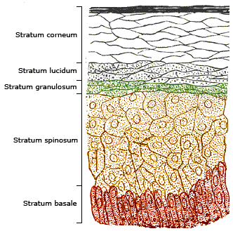
The skin consists of three main layers: epidermis, dermis, and hypodermis. The epidermis contains several strata, each with distinct functions.
Epidermis: Stratified squamous ET; layers include stratum corneum, lucidum, granulosum, spinosum, basale.
Dermis: Dense irregular CT; contains dermal papillae.
Hypodermis: Loose adipose CT; provides insulation and energy storage.
Accessory Structures: Arrector pili muscle, sebaceous gland, sweat gland, hair shaft.
The Skeletal System
Axial and Appendicular Skeletons
The skeleton is divided into axial (skull, vertebral column, ribcage) and appendicular (limbs, girdles) components.
Axial Skeleton: Includes skull, vertebrae, ribs, sternum.
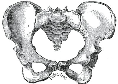
Appendicular Skeleton: Includes limbs, pectoral and pelvic girdles.
Bone Classification and Markings
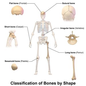
Bones are classified by shape: long, short, flat, irregular, sesamoid. Bone markings include foramen, condyle, epicondyle, meatus, process, fossa, tuberosity.
Long Bones: Femur, humerus
Short Bones: Carpals
Flat Bones: Skull, sternum
Irregular Bones: Vertebrae
Sesamoid Bones: Patella
The Muscular System
Skeletal Muscles and Actions
Skeletal muscles are responsible for voluntary movements. Each muscle has a specific action, often named for its function or location.
Flexion: Decreases angle between bones (e.g., biceps brachii flexes forearm).
Extension: Increases angle (e.g., triceps brachii extends forearm).
Abduction: Moves limb away from midline.
Adduction: Moves limb toward midline.
Dorsiflexion: Raises foot upward.
Plantarflexion: Points toes downward.
Rotation: Circular movement.
Circumduction: Cone-shaped movement.
The Nervous System
Brain and Spinal Cord Anatomy
The nervous system includes the brain and spinal cord, which are responsible for processing and transmitting information.
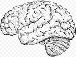
Brain: Cerebrum (lobes, hemispheres), cerebellum, diencephalon (thalamus, hypothalamus), brain stem (pons, medulla, midbrain).
Spinal Cord: White and gray matter, central canal, dorsal and ventral roots, spinal nerves.
The Cardiovascular System
Heart Anatomy and Blood Flow
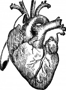
The heart is a muscular organ that pumps blood through the pulmonary and systemic circuits. Key structures include chambers, valves, and major vessels.
Chambers: Right/left atria, right/left ventricles
Valves: Tricuspid, bicuspid (mitral), pulmonary, aortic
Major Vessels: Aorta, pulmonary arteries/veins, vena cava
Blood Flow Pathway: SVC/IVC → right atrium → tricuspid valve → right ventricle → pulmonary valve → pulmonary arteries → lungs → pulmonary veins → left atrium → bicuspid valve → left ventricle → aortic valve → aorta
Blood Vessels
Arteries carry blood away from the heart; veins return blood to the heart. Major vessels are named for their location and function.
Aorta: Main artery
Vena Cava: Main vein
Radial, Brachial, Femoral: Named for nearby bones
The Respiratory System
Respiratory Anatomy and Pathways
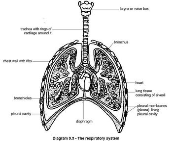
The respiratory system includes structures for gas exchange: nasal cavity, pharynx, larynx, trachea, bronchi, lungs.
Upper Respiratory Tract: Nasal cavity, oral cavity, pharynx, larynx
Lower Respiratory Tract: Trachea, bronchi, lungs
Pathway of Oxygen: Nares → nasal cavity → pharynx → larynx → trachea → bronchi → lungs → left atrium
Pathway of Carbon Dioxide: Pulmonary capillaries → lungs → bronchi → trachea → larynx → pharynx → nares
The Digestive System
Digestive Organs and Pathways
The digestive system breaks down food and absorbs nutrients. It includes the oral cavity, pharynx, esophagus, stomach, intestines, liver, pancreas, gallbladder.
Upper GI Tract: Mouth, pharynx, esophagus, stomach
Lower GI Tract: Small intestine (duodenum, jejunum, ileum), large intestine (cecum, colon, rectum, anus)
Accessory Organs: Liver, pancreas, gallbladder
Pathway of Food: Oral cavity → pharynx → esophagus → stomach → small intestine → large intestine → anus
The Urinary System
Urinary Anatomy and Pathways
The urinary system removes waste from the blood and regulates fluid balance. It includes kidneys, ureters, bladder, urethra.
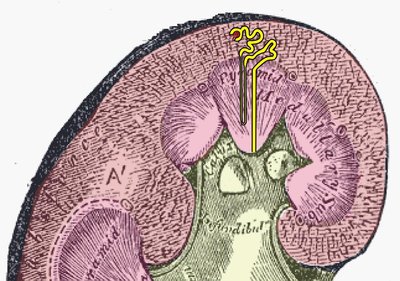
Kidney Regions: Cortex, medulla, pelvis
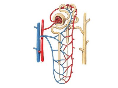
Nephron Structures: Glomerulus, Bowman's capsule, PCT, loop of Henle, DCT, collecting duct
Pathway of Urine: Glomerulus → Bowman's capsule → PCT → loop of Henle → DCT → collecting duct → renal pelvis → ureter → bladder → urethra
Abnormal Constituents of Urine
Abnormal substances in urine indicate possible disease or dysfunction.
Constituent | Symptoms | Causes |
|---|---|---|
Leukocytes | Frequent, painful urination, fever | Urinary tract infection |
Protein (albumin) | Swelling, fatigue | Exertion, pregnancy, heart failure, hypertension |
Erythrocytes | Painful urination, pink urine | Renal trauma, stones, infection, cancer |
Glucose | Frequent urination, hunger, thirst | Excess sugar intake, diabetes mellitus |

The Reproductive System
Male and Female Reproductive Anatomy
The reproductive system includes organs for producing gametes and supporting reproduction.
Male: Penis, scrotum, testes, epididymis, vas deferens, seminal vesicle, prostate, Cowper's gland, urethra
Female: Ovaries, uterine tubes, uterus (cervix, body, fundus), vagina, labia, clitoris
Reproductive Pathways
Sperm Pathway: Seminiferous tubules → epididymis → vas deferens → ejaculatory duct → urethra → outside
Oocyte Pathway: Ovary → uterine tube → uterus → vagina → delivery
Laboratory Safety Procedures
General and Personal Safety
Laboratory safety is essential for preventing accidents and injuries. Follow all written and verbal instructions, use appropriate PPE, and maintain a clean workspace.
Never eat, drink, or smoke in the lab.
Use equipment only if trained.
Wear lab coats, safety glasses, gloves as required.
Tie back long hair and secure loose clothing.
Clean up before leaving.
Know emergency procedures and locations of safety equipment.
Emergency Response
Read safety and fire alarm posters.
Know locations of fire extinguisher, eye wash, safety shower.
Notify instructor of any injury or spill.
Know evacuation procedures.
Call 911 and campus security in emergencies.
Additional info: Academic context and examples were added to clarify brief points and ensure completeness for exam preparation.
