 Back
Comprehensive Study Guide: Joints (Articulations) in Human Anatomy
Study Guide - Smart Notes
 Tailored notes based on your materials, expanded with key definitions, examples, and context.
Joints: An Overview
Definition and Function
Joints, also known as articulations, are the locations where two or more rigid elements of the skeleton meet. The Greek root arthro means joint. Joints enable the skeleton to resist crushing, tearing, and other forces, while also allowing for movement and flexibility.
Key Point: Joints are essential for both stability and mobility in the human body.
Key Point: The structure of a joint determines its function and range of movement.
Classification of Joints
Functional Classification
Joints are classified by the amount of movement they allow:
Synarthroses: Immovable joints; commonly found in the axial skeleton.
Amphiarthroses: Slightly movable joints; also common in the axial skeleton.
Diarthroses: Freely movable joints; typical of the appendicular skeleton (all synovial joints).
Structural Classification
Structural classification is based on the material binding the bones and the presence or absence of a joint cavity. The main types are:
Fibrous Joints: Bones connected by fibrous connective tissue; no joint cavity.
Cartilaginous Joints: Bones united by cartilage; lack a joint cavity.
Synovial Joints: Bones separated by a joint cavity filled with synovial fluid; most movable type.
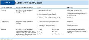
Summary Table of Joint Classes
Structural Class | Structural Characteristics | Types | Mobility |
|---|---|---|---|
Fibrous | Adjoining bones united by collagenic fibers | 1. Suture (short fibers) 2. Syndesmosis (longer fibers) 3. Gomphosis (periodontal ligament) | Immobile (synarthrosis) Slightly movable (amphiarthrosis) and immobile Immobile |
Cartilaginous | Adjoining bones united by cartilage | 1. Synchondrosis (hyaline cartilage) 2. Symphysis (fibrocartilage) | Immobile Slightly movable |
Synovial | Adjoining bones separated by a joint cavity, covered with articular cartilage, enclosed within an articular capsule lined with synovial membrane | 1. Plane 2. Hinge 3. Pivot 4. Condylar 5. Saddle 6. Ball-and-socket | Freely movable (diarthrosis); movements depend on design of joint |
Fibrous Joints
Types of Fibrous Joints
Fibrous joints are connected by fibrous connective tissue and do not have a joint cavity. Most are immovable or only slightly movable.
Sutures: Found only in the skull; bones are tightly bound by minimal fibrous tissue, allowing for bone growth during childhood. Eventually ossify to form synostoses.
Syndesmoses: Bones connected exclusively by ligaments; movement depends on fiber length. Example: tibiofibular joint (immovable), interosseous membrane between radius and ulna (freely movable).
Gomphoses: Peg-in-socket joint; example is the tooth in its socket, held by the periodontal ligament.
Cartilaginous Joints
Types of Cartilaginous Joints
Cartilaginous joints unite bones with cartilage and lack a joint cavity.
Synchondroses: Bones united by hyaline cartilage. Examples: epiphyseal plates in growing bones, joint between first rib and sternum.
Symphyses: Bones united by fibrocartilage, which resists tension and compression. Examples: intervertebral discs, pubic symphysis. These joints are slightly movable and provide strength with flexibility.
Synovial Joints
General Structure
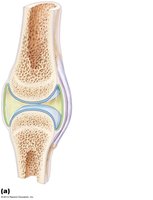
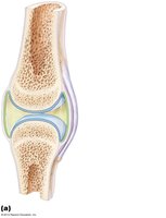
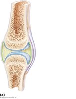
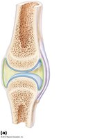
Synovial joints are the most movable type and are characterized by a fluid-filled joint cavity. All synovial joints are diarthroses (freely movable).
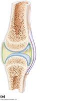
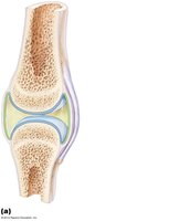
Articular Cartilage: Covers the ends of opposing bones with hyaline cartilage, absorbing compression.
Joint (Articular) Cavity: Unique to synovial joints; holds a small amount of synovial fluid.
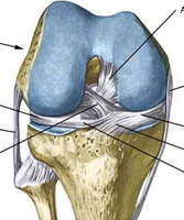
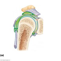
Articular Capsule: Encloses the joint cavity in a two-layered capsule: a fibrous layer (dense irregular connective tissue) and a synovial membrane (loose connective tissue).
Synovial Fluid: Viscous fluid, a filtrate of blood, containing glycoprotein molecules. Provides lubrication via "weeping lubrication"—fluid is squeezed in and out of articular cartilage under pressure.
Reinforcing Ligaments: Strengthen the joint; may be extracapsular (outside the capsule) or intracapsular (inside the capsule).
Sensory Nerves: Detect pain and monitor capsule stretch.
Blood Supply: Richly supplied; capillary beds produce synovial fluid.
Bursae and Tendon Sheaths
Bursae and tendon sheaths are associated with synovial joints and act as closed bags of lubricant to reduce friction between body elements.
Bursa: Flattened fibrous sac lined by a synovial membrane.
Tendon Sheath: An elongated bursa that wraps around a tendon.
Movements Allowed by Synovial Joints
Types of Movement
Synovial joints allow three basic types of movement:
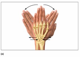
Gliding: One bone slides across the surface of another. Occurs in carpals, tarsals, and articular processes of vertebrae.
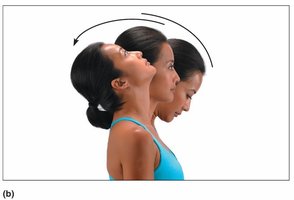
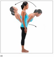
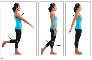
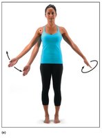
Angular Movement: Changes the angle between bones. Includes flexion, extension, abduction, adduction, and circumduction.
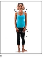
Rotation: Movement around a bone’s long axis. Occurs at the hip, shoulder, and between atlas and axis vertebrae.
Special Movements
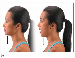
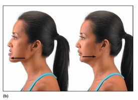
Elevation: Lifting a body part superiorly.
Depression: Moving a body part inferiorly.
Protraction: Moving a body part anteriorly.
Retraction: Moving a body part posteriorly.
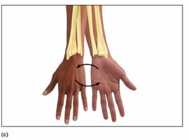
Supination: Rotating the forearm so the palm faces anteriorly.
Pronation: Rotating the forearm so the palm faces posteriorly.
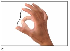
Opposition: Moving the thumb to touch the tips of other fingers.
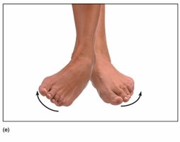
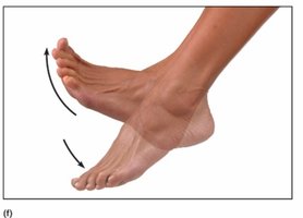
Inversion: Turning the sole of the foot medially.
Eversion: Turning the sole of the foot laterally.
Dorsiflexion: Lifting the foot so its superior surface approaches the shin.
Plantar Flexion: Depressing the foot, elevating the heel.
Synovial Joints Classified by Shape
Plane Joint
Articular surfaces are flat planes.
Short gliding movements; nonaxial.
Examples: intertarsal, intercarpal joints.
Hinge Joint
Cylindrical end of one bone fits into a trough on another.
Allows angular movement in one plane; uniaxial.
Examples: elbow, ankle, interphalangeal joints.
Pivot Joint
Rotating bone turns only around its long axis; uniaxial.
Examples: proximal radioulnar joint, atlantoaxial joint.
Condylar (Ellipsoid) Joint
Oval articular surfaces allow movement around two axes; biaxial.
Side to side (abduction/adduction) and back and forth (flexion/extension).
Examples: metacarpophalangeal (knuckle) joints, wrist joints.
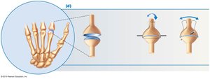
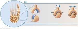
Saddle Joint
Each articular surface has concave and convex surfaces; biaxial.
Allows abduction/adduction and flexion/extension.
Example: first carpometacarpal joint (thumb).
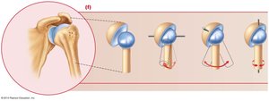
Ball-and-Socket Joint
Spherical head of one bone fits into round socket of another; multiaxial.
Allows movement in all axes.
Examples: shoulder and hip joints.
Disorders of Joints
Joint Injuries
Torn cartilage: Common injury to meniscus of knee joint.
Sprains: Ligaments of a reinforcing joint are stretched or torn.
Dislocation: Bones of a joint are forced out of alignment.
Inflammatory and Degenerative Conditions
Bursitis: Inflammation of a bursa due to injury or friction.
Tendonitis: Inflammation of a tendon.
Tenosynovitis: Inflammation of a tendon sheath.
Arthritis: Over 100 kinds of joint-damaging diseases.
Osteoarthritis: Most common type; "wear and tear" arthritis.
Rheumatoid arthritis: Chronic inflammatory disorder.
Gouty arthritis (gout): Uric acid buildup causes pain in joints.
Lyme disease: Inflammatory disease often resulting in joint pain.
Additional info: The notes above expand on the original content with academic context, definitions, and examples to ensure completeness and clarity for exam preparation.
