 Back
Comprehensive Study Guide: Skeletal, Muscular, Cardiovascular, and Lymphatic Systems
Study Guide - Smart Notes
 Tailored notes based on your materials, expanded with key definitions, examples, and context.
Skeletal System
Overview and Functions
The skeletal system provides the structural framework for the body and serves several essential functions:
Support: Framework for the body, e.g., vertebral column supports the trunk.
Protection: Shields vital organs, e.g., skull protects the brain, ribs protect the heart and lungs.
Movement: Bones act as levers, joints as fulcrums, and muscles pull on bones to produce movement.
Mineral & Lipid Storage: Stores calcium, phosphate, and fat (yellow marrow).
Blood Cell Formation (Hematopoiesis): Occurs in red bone marrow.
Calcium Homeostasis: Bone acts as a reservoir; regulated by hormones.
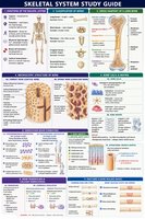
Bone Classification
Long Bones: Length > width (e.g., femur, humerus).
Short Bones: Cube-shaped (e.g., carpals, tarsals).
Flat Bones: Thin, often curved (e.g., skull, sternum).
Irregular Bones: Complex shapes (e.g., vertebrae, hip bones).
Sesamoid Bones: Form within tendons (e.g., patella).
Gross Anatomy of a Long Bone
Diaphysis: Shaft; compact bone around medullary cavity.
Epiphyses: Ends; mostly spongy bone with thin compact shell.
Metaphysis: Region between diaphysis & epiphysis; contains epiphyseal plate/line.
Articular Cartilage: Hyaline cartilage covering epiphyses.
Periosteum: Outer membrane (fibrous and osteogenic layers).
Endosteum: Inner lining of medullary cavity and trabeculae.
Microscopic Bone Structure
Compact Bone: Organized into osteons (Haversian systems) with central canals, lamellae, lacunae, canaliculi, and perforating canals.
Spongy Bone: Lattice-like trabeculae; spaces contain red marrow; no true osteons.
Bone Cells and Matrix
Osteogenic Cells: Stem cells; differentiate into osteoblasts.
Osteoblasts: Bone-forming; secrete osteoid.
Osteocytes: Mature cells in lacunae; maintain matrix.
Osteoclasts: Bone-resorbing; break down matrix.
Matrix: Organic (collagen, ground substance) for tensile strength; inorganic (hydroxyapatite) for hardness.
Ossification (Bone Formation)
Intramembranous Ossification: Forms flat bones directly from mesenchyme.
Endochondral Ossification: Forms most bones from a hyaline cartilage model.
Bone Growth and Remodeling
Longitudinal Growth: Occurs at epiphyseal plates via endochondral growth.
Appositional Growth: Increase in bone width by osteoblasts under periosteum.
Remodeling: Continuous deposition and resorption; regulated by mechanical stress and hormones (PTH, calcitonin).
Bone Fractures & Healing
Types: Simple, compound, comminuted, greenstick, compression.
Healing Steps: Hematoma formation, fibrocartilaginous callus, bony callus, remodeling.
Muscle Structure & Function
Skeletal Muscle Structure
Skeletal muscle is composed of bundles of muscle fibers (myofibers) wrapped in connective tissue sheaths. Each myofiber contains myofibrils, which are made of repeating units called sarcomeres.
Myofilaments: Actin (thin), myosin (thick), titin (elastic).
Sarcomere: Functional unit; extends from Z disc to Z disc.
Sarcolemma: Muscle cell membrane; conducts signals.
Sarcoplasmic Reticulum: Stores and releases Ca2+.
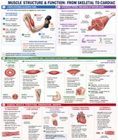
Sarcomere Bands & Contraction
I Band: Light; actin only; shortens during contraction.
A Band: Dark; myosin (with actin overlap); unchanged during contraction.
H Zone: Myosin only; disappears when contracted.
M Line: Center of sarcomere; anchors myosin.
Sliding Filament Mechanism
Explains muscle contraction at the sarcomere level: thin and thick filaments slide past each other.
Requires Ca2+ (activates actin) and ATP (activates myosin).
Nerve Supply & Neuromuscular Junction (NMJ)
Somatic Motor Neuron: Voluntary control of skeletal muscle.
Acetylcholine (ACh): Neurotransmitter at NMJ; excites muscle fiber.
NMJ Components: Axon terminal, synaptic cleft, sarcolemma.
Motor Units & Muscle Tone
Motor Unit: One motor neuron + all myofibers it innervates.
Muscle Tone: Constant, low-level contraction for posture and stability.
Muscle Growth, Atrophy, and Damage
Hypertrophy: Increase in cell size (more myofibrils/myofilaments).
Atrophy: Decrease in cell size (loss of cytoplasmic contents).
Fibrosis: Replacement of damaged muscle with connective tissue (scar).
Attachments, Levers, and Body Movements
Tendon: Connects muscle to bone.
Origin: Proximal/less mobile attachment.
Insertion: Distal/more mobile attachment.
Lever System: Bones (levers), joints (fulcrums), muscles (effort), load (resistance).
Group Actors in Movement
Agonist (Prime Mover): Main muscle for action (e.g., brachialis in elbow flexion).
Antagonist: Opposes action (e.g., triceps brachii).
Synergist: Aids agonist (e.g., biceps brachii).
Fixator: Stabilizes joints (e.g., rotator cuff muscles).
Power vs Range of Motion
More fascicles = more power; fewer fascicles = more range of motion (ROM).
Inverse relationship: ↑ Power → ↓ ROM; ↑ ROM → ↓ Power.
Cardiac Muscle: Structure, Innervation, Growth
Basic Properties
Location: Myocardium (heart wall).
Striated, involuntary, usually one central nucleus per cell.
Intercalated Discs: Contain gap junctions for rapid electrical impulse spread.
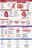
Nervous Innervation
Autonomic Nervous System: Controls heart rate automatically.
SA Node: Initiates impulse; AV Node: Relays to ventricles.
Coronary Circulation
Supplies O2-rich blood to heart muscle via coronary arteries.
Growth & Regeneration
No mitosis; growth by hypertrophy (cell enlargement).
Damage repaired by fibrosis (scar tissue).
Smooth Muscle: Structure, Innervation, Function
Basic Properties
No striations; fusiform cells with single central nucleus.
Involuntary; found in walls of hollow organs and arrector pili muscles.
Microscopic Anatomy
No sarcomeres; myofilaments arranged in a unique, non-striated pattern.
Gap junctions allow wave-like contractions.
Nerve Supply
Autonomic nervous system; varicosities form plexuses around cells.
Cardiovascular & Lymphatic Systems
Overview
Cardiovascular System: Heart, blood vessels, blood; transports O2, CO2, nutrients, wastes, hormones.
Lymphatic System: Returns excess interstitial fluid to blood; maintains blood volume and pressure.
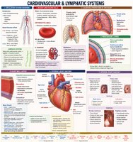
Blood & Red Blood Cells
Blood: Plasma (liquid), formed elements (RBCs, WBCs, platelets).
Erythrocytes: Biconcave, anucleate; transport O2 via hemoglobin.
Heart Location & Protection
Located in thoracic cavity, within mediastinum; protected by ribs, sternum, vertebrae.
Pericardium: Fibrous, parietal, visceral layers; pericardial cavity with serous fluid reduces friction.
Heart Wall Layers
Epicardium: Outer layer (visceral pericardium).
Myocardium: Cardiac muscle; thickest in left ventricle.
Endocardium: Inner lining; endothelium + loose CT.
External & Internal Heart Anatomy
Apex: Inferior tip; Base: Superior region.
Auricles: Flaps over atria; increase volume.
Sulci: Grooves housing coronary vessels.
Pectinate Muscles: Ridged atrial muscle.
Fossa Ovalis: Remnant of fetal foramen ovale.
Papillary Muscles & Chordae Tendineae: Stabilize AV valves.
Trabeculae Carneae: Irregular ventricular muscle ridges.
Heart Valves
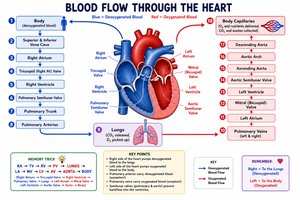
Atrioventricular (AV) Valves: Tricuspid (right), bicuspid/mitral (left); prevent backflow into atria.
Semilunar Valves: Pulmonary (right ventricle to pulmonary trunk), aortic (left ventricle to aorta); prevent backflow into ventricles.
Path of Blood Through the Heart
Right atrium (from SVC, IVC, coronary sinus)
Tricuspid valve → Right ventricle
Pulmonary semilunar valve → Pulmonary trunk → Pulmonary arteries
Lungs (gas exchange)
Pulmonary veins → Left atrium
Mitral valve → Left ventricle
Aortic semilunar valve → Aorta → Body
Systemic veins → SVC/IVC → Right atrium
Fibrous Skeleton & Conducting System
Fibrous Skeleton: Dense CT around valves; supports, anchors, and electrically separates atria from ventricles.
Conducting System: SA node → AV node → AV bundle → Bundle branches → Purkinje fibers; coordinates contraction.
Coronary Circulation
Supplies myocardium via right and left coronary arteries; returns via cardiac veins to coronary sinus.
Blood Vessels: Structure and Function
Blood Vessel Wall Layers (Tunics)
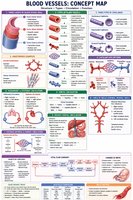
Tunica Intima: Endothelium (simple squamous); smooth blood flow.
Tunica Media: Smooth muscle + elastic fibers; controls diameter, blood pressure.
Tunica Externa (Adventitia): Connective tissue; anchors vessel.
Types of Blood Vessels
Elastic Arteries: Largest; stretch/recoil (e.g., aorta).
Muscular Arteries: Distribute blood to organs.
Arterioles: Control blood flow into capillaries.
Capillaries: Smallest; exchange of gases, nutrients, wastes.
Venules: Drain capillary beds.
Veins: Return blood to heart; have valves.
Types of Capillaries
Continuous: Least permeable; muscle, skin, CNS.
Fenestrated: Pores; kidneys, intestines, endocrine glands.
Sinusoid: Most permeable; liver, spleen, bone marrow.
Circulatory Pathways
Pulmonary Circulation: Right ventricle → lungs → left atrium; low pressure, short distance.
Systemic Circulation: Left ventricle → body → right atrium; high pressure, long distance.
Portal Circulation: Two capillary beds connected by a vein (e.g., hepatic portal system).
Vascular Disorders
Atherosclerosis: Plaque buildup in arteries.
Deep Vein Thrombosis (DVT): Blood clot in deep vein.
Varicose Veins: Swollen veins due to valve failure.
Aneurysm: Weak, bulging vessel wall.
Fetal Circulation
Bypasses lungs and liver via foramen ovale, ductus arteriosus, ductus venosus.
Changes at birth: closure of fetal shunts, formation of adult structures (e.g., fossa ovalis, ligamentum arteriosum).
Key Terms to Memorize
Artery, Vein, Capillary, Tunica Intima, Tunica Media, Tunica Externa, Elastic Artery, Muscular Artery, Arteriole, Venule, Capillary Bed, Precapillary Sphincter, Anastomosis, Pulmonary Circulation, Systemic Circulation, Hepatic Portal System, Atherosclerosis, Deep Vein Thrombosis, Aneurysm, Foramen Ovale
