 Back
BackComprehensive Study Guide: The Skeletal System
Study Guide - Smart Notes
 Tailored notes based on your materials, expanded with key definitions, examples, and context.
The Skeletal System
Overview of the Skeletal System
The skeletal system is a complex framework of bones and connective tissues that provides structure, protection, and movement for the human body. It is essential for supporting body weight, protecting internal organs, and facilitating movement through its joints and attachments to muscles.
Bone Tissue Type: Bone is a specialized connective tissue composed of cells, fibers, and ground substance.
Functions: Support, protection, movement, mineral storage (especially calcium and phosphate), and blood cell production (hematopoiesis).
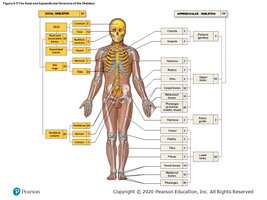
Divisions: The skeleton is divided into the axial skeleton (skull, vertebral column, rib cage) and the appendicular skeleton (limbs and girdles).
Total Bones: The adult human skeleton contains 206 bones.
Classification of Bones
Bones are classified by their shapes, which relate to their functions and locations in the body.
Long Bones: Longer than they are wide (e.g., femur, humerus).
Short Bones: Nearly equal in length and width (e.g., carpals, tarsals).
Flat Bones: Thin and broad (e.g., sternum, skull bones).
Irregular Bones: Complex shapes (e.g., vertebrae, pelvis).
Bone Surface Markings
Surface markings on bones serve as attachment points for muscles, tendons, and ligaments, and as passages for nerves and blood vessels.
Foramina: Openings for nerves and blood vessels.
Processes: Projections for muscle and ligament attachment.
Fossae: Depressions that may articulate with other bones.
Structure of a Long Bone
Long bones have distinct anatomical regions that contribute to their function and growth.
Diaphysis: The shaft, composed mainly of compact bone.
Epiphyses: The ends, consisting of spongy bone covered by compact bone.
Metaphysis: The region between diaphysis and epiphysis, containing the growth plate.
Medullary Cavity: Central cavity containing bone marrow.
Periosteum: Outer fibrous covering.
Endosteum: Lining of the medullary cavity.
Microscopic Structure of Bone
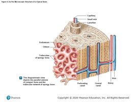
Bones are composed of compact and spongy tissue, each with specialized structures for strength and nutrient delivery.
Osteon (Haversian System): The structural unit of compact bone, consisting of concentric lamellae around a central canal.
Trabeculae: The lattice-like network in spongy bone.
Canaliculi: Small channels connecting osteocytes for nutrient exchange.
Bone Cells and Their Functions
Bone tissue is maintained and remodeled by three main cell types:
Osteocytes: Mature bone cells that maintain bone matrix.
Osteoblasts: Cells that build new bone matrix.
Osteoclasts: Cells that break down bone matrix for remodeling and calcium release.
Cartilage vs. Bone
Cartilage and bone are both connective tissues but differ in structure and function.
Cartilage: Flexible, avascular tissue found in joints, ear, nose, and respiratory tract.
Bone: Rigid, vascular tissue that forms the skeleton.
Growth of Bone
Bones grow in length and thickness through specific processes.
Appositional Growth: Increase in bone diameter by addition of new bone tissue at the surface.
Longitudinal Growth: Occurs at the epiphyseal plates (growth plates).
Fractures
Fractures are breaks in bones that can be classified by their appearance and cause. Recognizing fracture types is important for diagnosis and treatment.
The Axial Skeleton
Skull Bones and Sutures
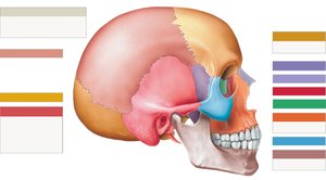
The skull is composed of several bones joined by immovable joints called sutures. These bones protect the brain and form the structure of the face.
Major Bones: Frontal, parietal, temporal, occipital, sphenoid, ethmoid, maxilla, mandible, zygomatic, nasal, lacrimal.
Sutures: Coronal (frontal to parietal), sagittal (between parietals), squamous (parietal to temporal), lambdoid (parietal to occipital).
Zygomatic Arch: Formed by the temporal process of the zygomatic bone and the zygomatic process of the temporal bone.
Vertebral Column
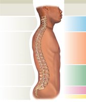
The vertebral column supports the body, protects the spinal cord, and allows flexible movement. It is divided into regions based on vertebral structure.
Cervical (7 vertebrae): Includes atlas (C1) and axis (C2), which support and rotate the head.
Thoracic (12 vertebrae): Articulate with ribs.
Lumbar (5 vertebrae): Support lower back.
Sacral and Coccygeal: Fused vertebrae forming the base of the spine.
Spinal Curves: Primary curves (thoracic, sacral) develop before birth; secondary curves (cervical, lumbar) develop after birth.
Rib Classification
Ribs are classified based on their attachment to the sternum.
True Ribs (1-7): Attach directly to the sternum.
False Ribs (8-12): Attach indirectly or not at all to the sternum.
Floating Ribs (11-12): Do not attach to the sternum.
The Appendicular Skeleton
Pectoral and Pelvic Girdles
The appendicular skeleton includes the bones of the limbs and the girdles that attach them to the axial skeleton.
Pectoral Girdle: Clavicle and scapula.
Pelvic Girdle: Hip bones (ilium, ischium, pubis).
Joints and Movements
Classification of Joints
Joints are classified by their structure and the amount of movement they allow.
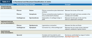
Functional Category | Structural Category and Type | Description | Example |
|---|---|---|---|
Synarthrosis (No Movement) | Fibrous: Suture | Fibrous connections plus interlocked surfaces | Between bones of the skull |
Fibrous: Gomphosis | Fibrous connection plus insertion in bony socket | Between teeth and jaws | |
Cartilaginous: Synchondrosis | Interposition of cartilage plate | Between first pair of ribs and sternum | |
Amphiarthrosis (Little Movement) | Fibrous: Syndesmosis | Ligamentous connection | Between tibia and fibula |
Cartilaginous: Symphysis | Connection by fibrocartilage pad | Between pubic bones | |
Diarthrosis (Free Movement) | Synovial | Complex joint bounded by joint capsule and containing synovial fluid | Shoulder, elbow, knee, etc. |
Synovial Joints
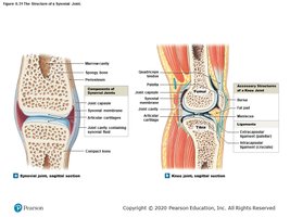
Synovial joints are the most movable type of joint, characterized by a joint capsule, synovial fluid, and articular cartilage.
Parts: Articular cartilage, joint cavity, synovial membrane, fibrous capsule, ligaments, tendons, bursae.
Function: Allow free movement and absorb shock.
Types of Synovial Joints
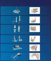
Synovial joints are classified by the shapes of their articulating surfaces and the movements they allow.
Plane (Gliding) Joint: Flat surfaces, slight movement (e.g., intercarpal joints).
Hinge Joint: Angular movement in one plane (e.g., elbow, knee).
Condylar (Ellipsoidal) Joint: Oval surface in depression, movement in two planes (e.g., wrist).
Saddle Joint: Concave and convex surfaces, movement in two planes plus circumduction (e.g., thumb).
Pivot Joint: Rotation in one plane (e.g., atlas and axis).
Ball-and-Socket Joint: Angular, rotational, and circumduction movement (e.g., shoulder, hip).
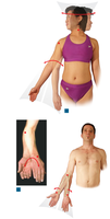
Movements of the Body
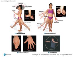
Body movements at synovial joints are described by specific terms based on the direction and nature of the movement.
Flexion/Extension: Decreasing/increasing the angle between bones.
Abduction/Adduction: Movement away from/toward the midline.
Rotation: Movement around a longitudinal axis.
Circumduction: Circular movement combining flexion, extension, abduction, and adduction.
Pronation/Supination: Rotational movement of the forearm.
Summary Table: Key Skeletal System Concepts
Concept | Key Points |
|---|---|
Bone Tissue | Specialized connective tissue; supports, protects, stores minerals, produces blood cells |
Bone Types | Long, short, flat, irregular |
Axial Skeleton | Skull, vertebral column, rib cage |
Appendicular Skeleton | Limbs, pectoral and pelvic girdles |
Joints | Classified by movement: synarthrosis, amphiarthrosis, diarthrosis (synovial) |
Movements | Flexion, extension, abduction, adduction, rotation, circumduction, pronation, supination |
Additional info: This guide expands on the provided review questions and images to create a comprehensive, exam-focused summary of the skeletal system, suitable for ANP college students.
