Back
BackComprehensive Study Notes for ANP College: Foundations, Cells, Genetics, and Nervous System
Study Guide - Smart Notes
 Tailored notes based on your materials, expanded with key definitions, examples, and context.
Lab Safety
Standard Safety Practices in Scientific Labs
Laboratory safety is essential to prevent accidents and ensure a safe working environment. The following protocols are standard in scientific settings:
Wear closed-toe shoes and long pants to protect the body from spills and hazards.
Personal Protective Equipment (PPE): Includes lab coat, masks, goggles, and gloves.
Hand hygiene: Wash hands before and after lab activities, even when gloves are worn.
No cell phones on bench tops to avoid contamination and distraction.
No food or drinks allowed in lab space to prevent ingestion of hazardous substances.
No inserting contacts or applying makeup during lab to avoid contamination.
Correct Hand-Washing Technique
Remove accessories (watches, jewelry) from fingers or wrists.
Wet hands under running water.
Apply soap and rub for 20 seconds; use a nail brush for nails.
Rinse hands thoroughly.
Dry hands with a clean paper towel; use it to turn off the tap.
Dispose of the paper towel in the trash.
Personal Protective Equipment (PPE)
PPE is used to minimize exposure to hazards. Common PPE includes lab coats, gloves, goggles, and masks, each serving to protect against specific risks such as chemical splashes, biological agents, or airborne particles.
Microscopy
Microscope Parts and Their Functions
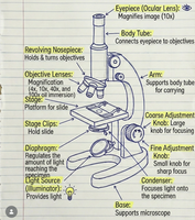
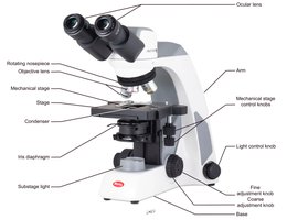
Understanding the parts of a microscope is crucial for proper use and maintenance. Key components include:
Eyepiece (Ocular Lens): Magnifies image, typically 10x.
Objective Lenses: Provide varying magnification (e.g., 4x, 10x, 40x).
Revolving Nosepiece: Holds and rotates objective lenses.
Stage: Platform for slide placement.
Stage Clips: Secure slides.
Diaphragm: Regulates light reaching the specimen.
Light Source: Provides illumination.
Coarse and Fine Adjustment Knobs: Focus the image.
Arm: Supports the body tube; used for carrying.
Base: Supports the microscope.
Condenser: Focuses light onto the specimen.
Calculating Total Magnification
Total magnification is determined by multiplying the power of the objective lens by the power of the ocular lens:
Formula:
Ocular lens power is typically 10x.
Example: If using a 40x objective lens, total magnification is .
Rules of Proper Microscope Use
Carry the microscope with one hand under the base and the other holding the arm.
Return the objective lens to scanning power before storing.
Use the coarse focus knob only with scanning and low-power objectives.
Do not touch the barrel; use the nosepiece to change objectives.
Report mechanical issues to the instructor.
Steps to View a Specimen
Obtain slide and microscope.
Turn on the microscope.
Place slide on stage and secure.
Rotate to scanning objective.
Use coarse knob to focus.
Use fine knob to sharpen image.
Center image with stage knobs.
Rotate to low-power objective.
Adjust brightness with diaphragm.
Use fine knob to sharpen focus.
Rotate to high-power objective.
Organization and Organ Systems
Levels of Biological Organization
Biological organization is hierarchical, from smallest to largest:
Atoms → Molecules → Cells → Tissues → Organs → Organ Systems → Organism
Anatomical Position
The anatomical position is a standard reference for describing body parts:
Body upright, arms at sides, palms forward, thumbs lateral.
Definitions: Anatomy and Physiology
Anatomy: Study of bodily structure and location.
Physiology: Study of normal functions of living organisms and their parts.
Body Cavities
Ventral Cavities: Thoracic, Abdominopelvic
Dorsal Cavities: Cranial, Vertebral
Major Organs in Organ Systems and Their Cavities
The following table summarizes the major organs in each system and their associated cavities:
Organ System | Organs | Cavity |
|---|---|---|
Integumentary | Skin, sweat glands, oil glands | None |
Renal/Urinary | Urinary bladder, ureters, kidneys | Abdominopelvic |
Lymphatic | Lymph nodes, vessels, red bone marrow, thymus, spleen, skin | Thoracic/Abdominal |
Muscular | Biceps, triceps, deltoid, gluteus maximus | None |
Cardiovascular | Heart, blood vessels | Thoracic |
Nervous | Brain, spinal cord | Cranial/Vertebral |
Respiratory | Lungs, trachea | Thoracic |
Digestive | Mouth, stomach, intestines, pancreas | Abdominopelvic |
Reproductive | Ovaries/testes, uterus, mammary glands, prostate | Abdominopelvic |
Endocrine | Pancreas, thyroid, thymus, ovaries/testes | Abdominopelvic |
Skeletal | Bones | None |
Chemical Composition of Cells
Dehydration Synthesis and Hydrolysis
Dehydration Synthesis: Removal of H2O to form polymers from monomers.
Hydrolysis: Addition of H2O to break polymers into monomers.
Positive and Negative Controls
Positive Control: Shows expected result when molecule is present.
Negative Control: Shows expected result when molecule is absent.
Simple vs. Complex Carbohydrates
Simple Carbohydrates: Monosaccharides and disaccharides (e.g., glucose, maltose).
Complex Carbohydrates: Polysaccharides (e.g., starch, glycogen).
Tests for Biomolecules
Biuret Test (Protein): Detects peptide bonds. Positive: violet/purple; negative: blue.
Benedict's Test (Simple Sugars): Detects glucose. Positive: color change (green, yellow, orange, red); negative: blue.
Iodine Test (Complex Sugars): Detects starch. Positive: blue/black; negative: yellow/brown.
Cell Structure and Function
Cell Organelles and Their Functions
Rough ER: Folds proteins into final shape.
Smooth ER: Processes lipids and carbohydrates.
Nucleus: Houses DNA.
Ribosome: Protein synthesis.
Mitochondria: Produces ATP.
Golgi Apparatus: Receives and delivers proteins and lipids.
Nucleolus: Ribosome production.
Lysosome: Breaks down waste.
Cell Membrane: Controls entry and exit of substances.
Diffusion Across the Cell Membrane
Plasma Membrane: Selectively permeable, composed of phospholipid bilayer and embedded proteins.
Simple Diffusion: Molecules move from high to low concentration without proteins.
Facilitated Diffusion: Requires membrane proteins; each protein is specific to a molecule type.
Genetics
Key Genetic Concepts
Chromosome: Collection of DNA; humans have 23 pairs.
Somatic Cells: Diploid (46 chromosomes).
Gamete Cells: Haploid (23 chromosomes).
Autosomes: First 22 pairs; homologous.
Sex Chromosomes: 23rd pair; not homologous.
Gene: DNA sequence coding for proteins.
Allele: Alternate versions of a gene; dominant (T) or recessive (t).
Genotype vs. Phenotype
Genotype: Combination of alleles (TT, tt, Tt).
Phenotype: Physical expression (hair color, eye color, etc.).
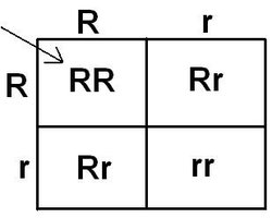
Punnett Square: Probability of Inheriting Traits
Punnett squares are used to predict the probability of offspring inheriting specific traits based on parental genotypes.
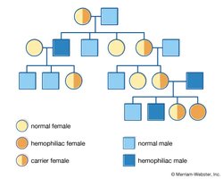
Pedigree Chart: Inheritance Patterns
Pedigree charts help determine inheritance patterns in families, such as autosomal dominant, autosomal recessive, or sex-linked traits.
Nervous System
Types of Cells in Nervous Tissue
Neuron: Transmits impulses and information.
Neuroglia: Supports neurons (includes Schwann cells, astrocytes, oligodendrocytes).
Structure of a Neuron
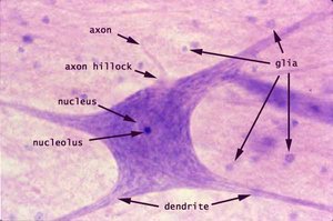
Neurons have distinct parts visible under a microscope:
Axon: Conducts impulses away from cell body.
Dendrite: Receives signals.
Nucleus: Contains genetic material.
Nucleolus: Ribosome production.
Axon Hillock: Initiates action potential.
Glia: Support cells.
Parts of the Brain and Their Functions
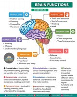
The brain is divided into regions, each with specialized functions:
Frontal Lobe: Problem solving, planning, personality, movement.
Parietal Lobe: Touch, spatial awareness, reading.
Temporal Lobe: Memory, hearing, language.
Occipital Lobe: Vision, color recognition.
Cerebellum: Balance, fine motor control.
Brainstem: Basic life functions.
Anatomy of the Spinal Cord
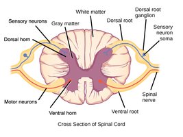
The spinal cord is organized into gray and white matter, with dorsal and ventral horns, and roots for sensory and motor neurons.
Gray Matter: Contains neuron cell bodies.
White Matter: Contains myelinated axons.
Dorsal Root: Sensory input.
Ventral Root: Motor output.
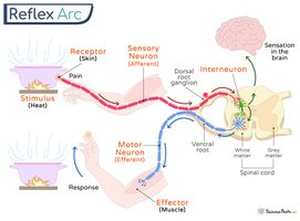
Components of a Reflex Arc
A reflex arc is the pathway by which a reflex occurs, involving:
Receptor: Detects stimulus.
Sensory Neuron: Transmits signal to CNS.
Interneuron: Processes signal in CNS.
Motor Neuron: Sends response to effector.
Effector: Muscle or gland that responds.