 Back
Comprehensive Study Notes for Human Anatomy & Physiology Laboratory (BIO 112)
Study Guide - Smart Notes
 Tailored notes based on your materials, expanded with key definitions, examples, and context.
Introduction to Human Anatomy & Physiology Laboratory
Laboratory Safety Procedures and Protocols
Laboratory safety is essential for preventing accidents and ensuring a productive learning environment. Students must adhere to all written and verbal safety instructions, use common sense, and be aware of emergency procedures.
Personal Safety: No eating, drinking, or smoking in the lab. Use equipment only if trained. Wear appropriate PPE (lab coats, safety glasses, gloves) as instructed.
Emergency Response: Know the location and use of fire extinguishers, eye wash stations, and safety showers. Report all injuries or accidents immediately. Be familiar with evacuation procedures and first aid kit locations.
General Conduct: Horseplay is strictly prohibited. Keep workspaces clean and organized. Wash hands before leaving the lab.
The Human Body: An Orientation
Anatomical Position and Its Importance
The anatomical position is a standardized posture used as a reference in describing the locations and relationships of body parts. In this position, the body stands upright, facing forward, with arms at the sides and palms facing forward. This consistency allows for clear communication in anatomy.
Why it is used: Provides a universal frame of reference for anatomical terminology and avoids confusion.
Directional Terms
Directional terms describe the positions of structures relative to other structures or locations in the body.
Superior: Toward the head
Inferior: Away from the head
Anterior (Ventral): Toward the front/belly side
Posterior (Dorsal): Toward the back
Medial: Toward the midline
Lateral: Away from the midline
Proximal: Closer to the point of attachment
Distal: Farther from the point of attachment
Superficial: Toward the surface
Deep: Away from the surface, toward the interior
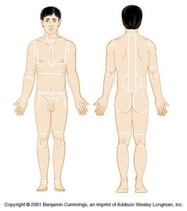
Anatomical Landmarks (Body Regions)
Body regions are specific areas used to describe locations on the body. Proper terminology is essential for accuracy in identification.
Examples: Brachial (arm), Femoral (thigh), Gluteal (buttocks), Cervical (neck), Abdominal (abdomen), etc.
Body Planes and Sections
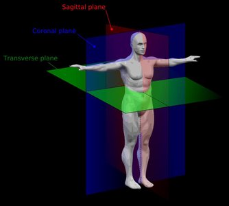
Body planes are imaginary lines used to divide the body into sections for anatomical study.
Sagittal Plane: Divides the body into left and right portions
Midsagittal Plane: Divides the body into equal left and right halves
Transverse (Horizontal) Plane: Divides the body into superior and inferior portions
Coronal (Frontal) Plane: Divides the body into anterior and posterior portions
Body Cavities
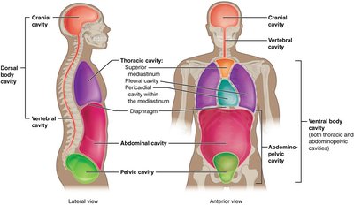
The body contains several major cavities that house and protect organs.
Ventral Body Cavity: Located on the anterior side; includes the thoracic and abdominopelvic cavities.
Thoracic Cavity: Contains the heart (pericardial cavity), lungs (pleural cavities), and mediastinum (central compartment).
Abdominopelvic Cavity: Subdivided into abdominal (digestive organs) and pelvic (bladder, reproductive organs) cavities.
Dorsal Body Cavity: Located on the posterior side; includes the cranial cavity (brain) and vertebral cavity (spinal cord).
Cells and Mitosis
Cellular Organelles and Their Functions
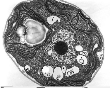
Cells contain specialized structures called organelles, each with a unique function essential for cell survival and activity.
Organelle | Function |
|---|---|
Plasma Membrane | Controls passage of materials into and out of the cell |
Cytoplasm | Contains organelles and dissolved solutes |
Nucleus | Contains DNA; controls cell activities and mitosis |
Rough Endoplasmic Reticulum (RER) | Protein synthesis (with ribosomes) |
Smooth Endoplasmic Reticulum (SER) | Lipid and membrane production |
Golgi Apparatus | Modifies and packages proteins for export |
Mitochondria | ATP production via aerobic respiration |
Centrioles | Form spindle fibers during mitosis |
The Cell Cycle and Mitosis
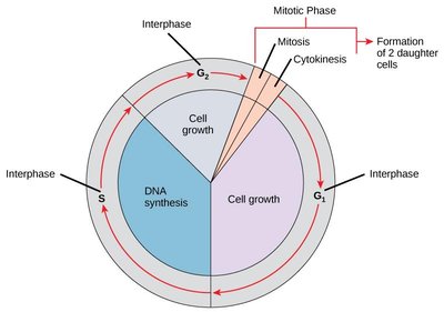
The cell cycle consists of interphase (cell growth and DNA replication) and mitosis (cell division). Mitosis is divided into four phases: prophase, metaphase, anaphase, and telophase, followed by cytokinesis.
Interphase: Cell grows, DNA replicates (G1, S, G2 phases)
Prophase: Chromatin condenses, nuclear envelope dissolves, spindle forms
Metaphase: Chromosomes align at the cell's equator
Anaphase: Sister chromatids separate to opposite poles
Telophase: Nuclear envelope reforms, chromosomes decondense
Cytokinesis: Division of cytoplasm, forming two daughter cells
Tissues and Integumentary System
Histology: The Study of Tissues
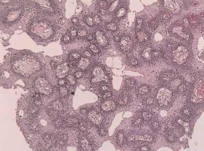
Histology is the study of tissues. Tissues are groups of similar cells performing a common function. There are four primary tissue types: epithelial, connective, muscle, and nervous.
Epithelial Tissue (ET): Covers surfaces, lines cavities, forms glands
Connective Tissue (CT): Supports, binds, and protects organs
Muscle Tissue (MT): Produces movement
Nervous Tissue (NT): Conducts impulses for communication
Major Tissue Types and Examples
General Type | Specific Type | Location | Function |
|---|---|---|---|
Epithelial | Simple squamous | Lung alveoli, capillaries | Diffusion |
Epithelial | Simple cuboidal | Kidney tubules, glands | Secretion, absorption |
Epithelial | Simple columnar | Digestive tract lining | Secretion, absorption |
Epithelial | Stratified squamous | Epidermis, mouth lining | Protection from abrasion |
Connective | Loose areolar | Deep to all ET | Nourishes ET via blood vessels |
Connective | Loose adipose | Hypodermis, breasts | Insulation, energy storage |
Connective | Dense regular | Tendons, ligaments | Strong attachment in one direction |
Connective | Hyaline cartilage | Ends of long bones | Flexible support, reduces friction |
Connective | Osseous (bone) | Skeleton | Support, movement |
Connective | Blood | Cardiovascular system | Transport |
Muscle | Cardiac | Heart | Pumps blood |
Muscle | Skeletal | Attached to skeleton | Voluntary movement |
Muscle | Smooth | Walls of hollow organs | Involuntary movement |
Nervous | Nervous tissue | Brain, spinal cord | Sensory input, control |
Skin Structure and Layers
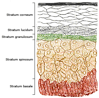
The skin is composed of three main layers: epidermis, dermis, and hypodermis. The epidermis consists of several strata, each with distinct characteristics.
Epidermis: Stratified squamous ET; layers include stratum corneum, lucidum, granulosum, spinosum, basale
Dermis: Dense irregular CT; contains dermal papillae
Hypodermis: Loose adipose CT; provides insulation and energy storage
Accessory Structures: Hair shaft, arrector pili muscle, sebaceous gland, sweat gland and pore
The Skeletal System
Axial and Appendicular Skeletons
The human skeleton is divided into two main parts:
Axial Skeleton: Skull, vertebral column, rib cage
Appendicular Skeleton: Limbs and girdles (shoulder and pelvic)
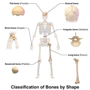
Classification of Bones by Shape
Long bones: Longer than wide (e.g., femur, humerus)
Short bones: Cube-shaped (e.g., carpals, tarsals)
Flat bones: Thin, flattened, often curved (e.g., sternum, skull bones)
Irregular bones: Complex shapes (e.g., vertebrae, pelvic bones)
Bone Markings
Bones have specific markings that serve as attachment points for muscles, passageways for nerves and blood vessels, and articulation points for joints.
Foramen: Hole for passage of vessels/nerves
Condyle: Rounded projection for articulation
Process: Projection for muscle/ligament attachment
Fossa: Shallow depression
Tuberosity: Roughened area for muscle attachment
The Nervous System
Major Structures of the Brain and Spinal Cord
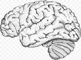
The nervous system is divided into the central nervous system (CNS) and peripheral nervous system (PNS). The CNS includes the brain and spinal cord.
Brain: Cerebrum (frontal, parietal, occipital, temporal lobes), cerebellum, diencephalon (thalamus, hypothalamus, epithalamus), brain stem (midbrain, pons, medulla oblongata)
Spinal Cord: White matter, gray matter, central canal, dorsal and ventral roots, spinal nerves
The Cardiovascular System
Heart Anatomy and Blood Flow
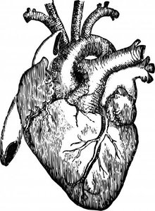
The heart is a muscular organ with four chambers (right/left atria, right/left ventricles) and valves that ensure unidirectional blood flow. Blood circulates through the pulmonary (lungs) and systemic (body) circuits.
Major Structures: Apex, aorta, atria, ventricles, valves (tricuspid, bicuspid/mitral, semilunar), septum, myocardium, endocardium, epicardium
Blood Flow Pathway: Superior/inferior vena cava → right atrium → tricuspid valve → right ventricle → pulmonary valve → pulmonary arteries → lungs → pulmonary veins → left atrium → bicuspid valve → left ventricle → aortic valve → aorta
The Respiratory System
Major Structures and Pathway of Air
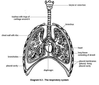
The respiratory system includes the nasal cavity, pharynx, larynx, trachea, bronchi, and lungs. Oxygen enters through the nares, passes through the conducting airways, and reaches the alveoli for gas exchange.
Upper Respiratory Tract: Nasal cavity, pharynx, larynx
Lower Respiratory Tract: Trachea, bronchi, lungs
Pathway of Oxygen: Nares → nasal cavity → pharynx → larynx → trachea → bronchi → bronchioles → alveoli → pulmonary capillaries → left atrium
The Urinary System
Major Organs and Pathway of Urine
The urinary system filters blood, removes waste, and regulates fluid balance. Major organs include the kidneys, ureters, urinary bladder, and urethra.
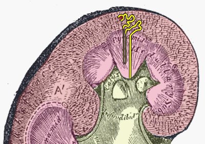
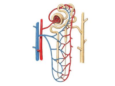
Kidney Structure: Renal cortex, renal medulla, renal pelvis, nephrons (glomerulus, tubules)
Pathway of Urine: Glomerulus → Bowman's capsule → proximal convoluted tubule → loop of Henle → distal convoluted tubule → collecting duct → renal pelvis → ureter → bladder → urethra
Abnormal Constituents of Urine
Constituent | Symptoms | Possible Causes |
|---|---|---|
Leukocytes | Painful urination, fever | Urinary tract infection |
Protein (albumin) | Swelling, fatigue | Exertion, pregnancy, heart failure, hypertension |
Erythrocytes | Pain, pink urine | Renal trauma, stones, infection, cancer |
Glucose | Frequent urination, thirst | High sugar intake, diabetes mellitus |

The Reproductive System
Male and Female Reproductive Anatomy
The reproductive system includes organs responsible for producing gametes and supporting fertilization and development.
Male Structures: Penis, scrotum, testes, epididymis, vas deferens, seminal vesicle, prostate gland, Cowper's gland, urethra
Female Structures: Ovaries, uterine tubes (fallopian tubes), uterus (cervix, body, fundus), vagina, labia majora/minora, clitoris
Pathways
Sperm Pathway: Seminiferous tubules → epididymis → vas deferens → ejaculatory duct → urethra → external environment
Oocyte Pathway: Ovary → uterine tube → uterus → (if fertilized) implantation and development → delivery
Study Strategies and Exam Preparation
Practice identifying structures on models and diagrams.
Memorize terminology, especially for anatomical regions, planes, and tissue types.
Understand the flow of substances (blood, air, urine, food, gametes) through organ systems.
Review tables and diagrams for quick recall during exams.
