Back
Comprehensive Study Notes for Human Anatomy & Physiology Lab (BIOL 2201)
Study Guide - Smart Notes
 Tailored notes based on your materials, expanded with key definitions, examples, and context.
Body Organization and Terminology
Anatomical Directions
Understanding anatomical directions is essential for accurately describing the locations and relationships of structures in the human body.
Superior: Toward the head or upper part of a structure.
Inferior: Away from the head or toward the lower part of a structure.
Anterior (Ventral): Toward the front of the body.
Posterior (Dorsal): Toward the back of the body.
Medial: Toward the midline of the body.
Lateral: Away from the midline of the body.
Proximal: Closer to the origin of a body part or the point of attachment.
Distal: Farther from the origin of a body part or the point of attachment.
Planes of the Body
Body planes are imaginary lines used to divide the body into sections for anatomical study.
Sagittal Section: Divides the body into right and left parts.
Transverse (Horizontal) Section: Divides the body into superior and inferior parts.
Frontal (Coronal) Section: Divides the body into anterior and posterior parts.
Abdominal Quadrants
The abdomen is divided into four quadrants for clinical and anatomical reference:
Right Upper Quadrant (RUQ)
Left Upper Quadrant (LUQ)
Right Lower Quadrant (RLQ)
Left Lower Quadrant (LLQ)
Body Cavities and Associated Membranes
The body contains several major cavities, each lined with membranes that protect and support internal organs.
Cranial Cavity: Contains the brain.
Thoracic Cavity: Contains the heart and lungs; includes the parietal pleura (lining the cavity), visceral pleura (covering the lungs), and the pleural cavity (space between pleurae).
Mediastinum: Central compartment of the thoracic cavity.
Diaphragm: Muscle separating thoracic and abdominopelvic cavities.
Abdominopelvic Cavity: Subdivided into the abdominal cavity (digestive organs) and pelvic cavity (urinary bladder, reproductive organs).
Peritoneum: Serous membrane of the abdominopelvic cavity, with parietal and visceral layers and the peritoneal cavity between them.
Tissues and Histology
Major Tissue Types
The human body is composed of four primary tissue types, each with distinct functions and characteristics.
Epithelial Tissue: Covers body surfaces and lines cavities; functions in protection, absorption, and secretion.
Connective Tissue: Supports, binds, and protects organs; includes bone, blood, cartilage, and adipose tissue.
Muscle Tissue: Responsible for movement; includes skeletal, cardiac, and smooth muscle.
Nervous Tissue: Conducts electrical impulses; found in the brain, spinal cord, and nerves.
Integumentary System (Skin)
Skin Structure
The skin is the largest organ of the body and consists of three main layers:
Epidermis: Outermost layer; includes the stratum corneum (dead, keratinized cells) and stratum basale (mitotically active cells).
Dermis: Middle layer; contains the papillary layer (areolar connective tissue) and reticular layer (dense irregular connective tissue), as well as structures like sweat glands, sebaceous glands, hair follicles, and sensory receptors.
Hypodermis (Subcutaneous Layer): Deepest layer; composed mainly of adipose tissue for insulation and energy storage.
Microscopic Features
Stratified Squamous Epithelium: Found in the epidermis, provides protection.
Meissner’s (Tactile) Corpuscle: Detects light touch.
Pacinian (Lamellar) Corpuscle: Detects deep pressure and vibration.
Skeletal System
Bone Structure
Bones are complex organs composed of several tissues and have various structural features:
Compact Bone: Dense outer layer providing strength.
Spongy Bone: Porous inner layer containing red marrow.
Epiphysis: End of a long bone.
Diaphysis: Shaft of a long bone.
Medullary Canal: Central cavity containing yellow marrow.
Microscopic Structure (Osteon Model)
Osteon: Structural unit of compact bone.
Central Canal: Contains blood vessels and nerves.
Lamellae: Concentric rings of bone matrix.
Osteocytes in Lacunae: Mature bone cells in small spaces.
Canaliculi: Tiny canals connecting osteocytes.
Axial and Appendicular Skeleton
Axial Skeleton: Skull, vertebral column, thoracic cage.
Appendicular Skeleton: Limbs and girdles (pectoral and pelvic).
Joints
Classification of Joints
Joints are classified based on their structure and function:
Fibrous Joints (Synarthroses): Immovable; e.g., sutures of the skull.
Cartilaginous Joints (Amphiarthroses): Slightly movable; e.g., intervertebral discs, pubic symphysis.
Synovial Joints (Diarthroses): Freely movable; e.g., shoulder, hip, knee.
Muscle Tissue and Muscles
Types of Muscle Tissue
Skeletal Muscle: Voluntary, striated, multinucleated.
Cardiac Muscle: Involuntary, striated, intercalated discs.
Smooth Muscle: Involuntary, non-striated, spindle-shaped cells.
Major Muscle Groups
Head and Neck: Frontalis, occipitalis, orbicularis oculi, masseter, temporalis, sternocleidomastoid.
Chest and Back: Trapezius, latissimus dorsi, pectoralis major/minor, deltoid, rotator cuff muscles.
Arm: Biceps brachii, triceps brachii, brachialis, brachioradialis.
Abdominal Wall: External oblique, rectus abdominis.
Hip and Leg: Gluteus maximus, quadriceps femoris group, hamstrings, gastrocnemius, tibialis anterior.
Nervous System
Fundamentals of Nervous Tissue
Neuron: Functional unit; consists of cell body, dendrites, axon.
Neuroglia (Glial Cells): Support and protect neurons.
Myelin Sheath: Insulates axons, increases conduction speed.
Node of Ranvier: Gaps in myelin sheath for saltatory conduction.
Central and Peripheral Nervous Systems
Central Nervous System (CNS): Brain and spinal cord.
Peripheral Nervous System (PNS): Cranial and spinal nerves, ganglia.
Spinal Cord: White matter (outer), gray matter (inner "H" shape), meninges (dura mater, arachnoid, pia mater).
Plexuses: Cervical, brachial, lumbar, sacral.
Special Senses
Major Sensory Structures
Skin: Meissner's corpuscle (touch), Pacinian corpuscle (pressure).
Tongue: Papillae, taste buds, gustatory cells.
Eye: Cornea, lens, retina, optic nerve, rods and cones.
Ear: Auricle, tympanic membrane, ossicles, cochlea, vestibulocochlear nerve.
Endocrine System
Major Endocrine Glands
Pituitary Gland (Hypophysis): Master gland controlling other endocrine glands.
Thyroid Gland: Regulates metabolism.
Adrenal Glands: Produce hormones for stress response.
Pancreas: Regulates blood glucose via insulin and glucagon.
Blood and Heart
Blood Components
Erythrocytes (RBCs): Transport oxygen.
Leukocytes (WBCs): Immune defense; classified as granulocytes (neutrophils, eosinophils, basophils) and agranulocytes (monocytes, lymphocytes).
Platelets: Blood clotting.
Heart Structure
Chambers: Right/left atria, right/left ventricles.
Valves: Tricuspid, mitral (bicuspid), pulmonary semilunar, aortic semilunar.
Major Vessels: Aorta, vena cava, pulmonary arteries/veins.
Blood Vessels
Major Arteries and Veins
Arteries: Carry blood away from the heart (e.g., aorta, carotid, femoral).
Veins: Return blood to the heart (e.g., vena cava, jugular, saphenous).
Immune System
Lymphatic Structures
Lymph Nodes: Filter lymph (cervical, axillary, inguinal, submandibular).
Spleen: Filters blood, immune response.
Tonsils: Immune surveillance in the pharynx.
Lymphatic Vessels: Transport lymph throughout the body.
Respiratory System
Major Structures
Nasal Cavity, Pharynx, Larynx: Air passageways; larynx contains vocal cords.
Trachea: Windpipe supported by hyaline cartilage.
Bronchi and Lungs: Branching airways ending in alveoli for gas exchange.
Pleura: Parietal and visceral layers with pleural cavity between.
Digestive System
Major Organs and Structures
Oral Cavity: Teeth, tongue, salivary glands.
Esophagus, Stomach, Small and Large Intestine: Digest and absorb nutrients.
Liver, Gallbladder, Pancreas: Accessory organs aiding digestion.
Intestinal Wall: Mucosa, submucosa, muscularis, serosa, villi, lacteals.
Urinary System
Major Structures
Kidneys: Filter blood, produce urine.
Ureters: Transport urine to bladder.
Urinary Bladder: Stores urine.
Urethra: Conducts urine out of the body.
Nephron: Functional unit of the kidney; includes glomerulus, tubules, collecting duct.
Reproductive System
Male and Female Structures
Male: Testes, epididymis, vas deferens, seminal vesicles, prostate, penis.
Female: Ovaries, uterine tubes, uterus, cervix, vagina, vulva.
Uterus: Fundus, body, cervix, endometrium, myometrium.
Human Body Proportions
Vitruvian Man
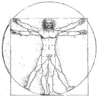
The Vitruvian Man is a famous drawing by Leonardo da Vinci, illustrating ideal human body proportions based on geometric principles. It is often used as a symbol of the study of anatomy and the harmony of the human form.