 Back
Comprehensive Study Notes: Joints (Articulations) in Human Anatomy & Physiology
Study Guide - Smart Notes
 Tailored notes based on your materials, expanded with key definitions, examples, and context.
Joints (Articulations)
Introduction to Joints
Joints, also known as articulations, are the anatomical locations where two bones connect. Because bones are rigid and relatively inflexible, movement in the body can only occur at joints. Some joints allow for movement, while others do not.
Definition: A joint is the site where two or more bones meet.
Function: Joints provide mobility and hold the skeleton together.
Types: Some joints are immovable, while others permit varying degrees of movement.
Classification of Joints
Joints are classified in two main ways: structurally (by the material binding the bones and the presence of a joint cavity) and functionally (by the amount of movement allowed).
Structural Classification: Based on the material connecting the bones and the presence or absence of a joint cavity.
Functional Classification: Based on the degree of movement permitted by the joint.
Structural Classification of Joints
Fibrous Joints: Bones are joined by fibrous connective tissue; no joint cavity is present.
Cartilaginous Joints: Bones are joined by cartilage; no joint cavity is present.
Bony Fusion (Synostosis): Bones fuse to form a single bony structure.
Synovial Joints: Bones are separated by a fluid-filled joint cavity.
Functional Classification of Joints
Synarthrosis: Immovable joint (e.g., sutures of the skull).
Amphiarthrosis: Slightly movable joint (e.g., pubic symphysis, intervertebral discs).
Diarthrosis: Freely movable joint (e.g., shoulder, hip, knee).
Types of Synarthroses (Immovable Joints)
Suture: Fibrous joint found between bones of the skull.
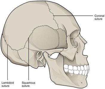
Gomphosis: Fibrous joint binding teeth to their sockets via the periodontal ligament.
Synchondrosis: Cartilaginous joint where bones are united by hyaline cartilage (e.g., first sternocostal joint).
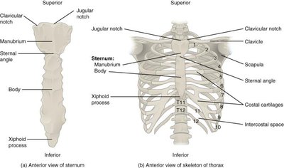
Synostosis: Bony fusion where two bones fuse and the boundary disappears (e.g., epiphyseal lines of long bones).
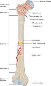
Types of Amphiarthroses (Slightly Movable Joints)
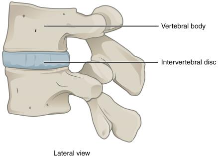
Symphysis: Cartilaginous joint where bones are separated by a pad of fibrocartilage (e.g., pubic symphysis, intervertebral discs).
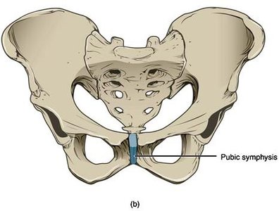
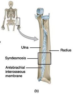
Syndesmosis: Fibrous joint where bones are connected by a ligament or interosseous membrane (e.g., between radius and ulna, tibia and fibula).
Types of Diarthroses (Freely Movable Joints / Synovial Joints)
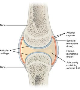
Diarthroses, or synovial joints, permit a wide range of motion and are characterized by a fluid-filled joint cavity.
Examples: Shoulder, elbow, hip, wrist, and ankle joints.
Structure of Synovial Joints
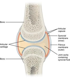
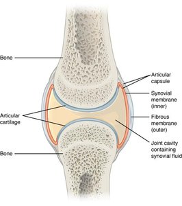
Articular Capsule: Encloses the joint cavity and is composed of fibrous connective tissue.
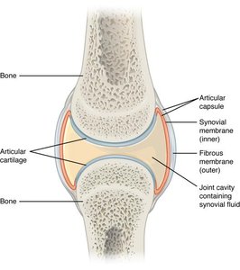
Synovial Membrane: Lines the inner surface of the capsule and secretes synovial fluid.
Synovial Fluid: Lubricates the joint, nourishes articular cartilage, and absorbs shock.
Articular Cartilage: Covers the articulating surfaces of bones, reducing friction and absorbing shock.
Accessory Ligaments: Support, strengthen, and reinforce synovial joints. Can be extracapsular (outside the capsule) or intracapsular (inside the capsule).
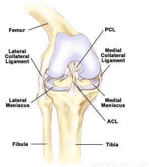
Meniscus (plural: menisci): Pads of fibrocartilage that improve fit between bones, absorb shock, and distribute weight (e.g., knee joint).
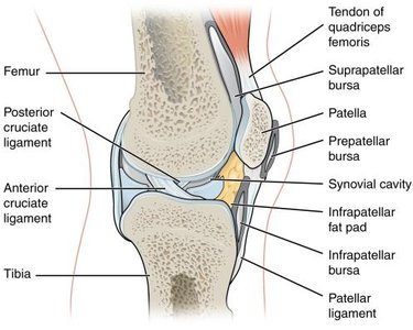
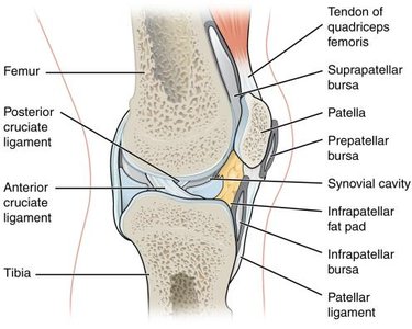
Bursa (plural: bursae): Small sacs filled with synovial fluid that reduce friction between moving structures (e.g., between skin and patella, or tendon and bone).
Classification of Synovial Joints by Axis of Motion
Uniaxial Joints: Allow movement in one plane (e.g., elbow, ankle).
Biaxial Joints: Allow movement in two planes (e.g., wrist, knuckle joints).
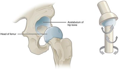
Triaxial Joints: Allow movement in three planes (e.g., hip, shoulder).
Types of Synovial Joints by Shape
Pivot Joint: Rounded bone fits into a ring, allowing rotation (e.g., proximal radioulnar joint).
Hinge Joint: Convex surface fits into concave surface, allowing flexion and extension (e.g., elbow, knee).
Condyloid Joint: Oval articular surface fits into a depression, allowing movement in two planes (e.g., wrist).
Plane (Planar) Joint: Flat surfaces allow bones to slide past each other (e.g., intercarpal joints).
Saddle Joint: Each articular surface has both concave and convex areas, allowing biaxial movement (e.g., thumb carpometacarpal joint).
Ball-and-Socket Joint: Spherical head fits into a cup-like socket, allowing the greatest range of motion (e.g., shoulder, hip).
Movements at Synovial Joints
Flexion: Decreases the angle between two bones (e.g., bending the elbow).
Extension: Increases the angle between two bones (e.g., straightening the knee).
Hyperextension: Extension beyond the anatomical position.
Abduction: Movement away from the midline.
Adduction: Movement toward the midline.
Circumduction: Circular movement of a limb.
Rotation: Movement around a bone's long axis (e.g., head rotation at the atlas-axis joint).
Supination and Pronation: Movements of the forearm; supination turns the palm anteriorly, pronation turns it posteriorly.
Dorsiflexion and Plantar Flexion: Movements at the ankle; dorsiflexion lifts the foot upward, plantar flexion points the toes downward.
Protraction and Retraction: Anterior and posterior movements in the horizontal plane (e.g., scapula, mandible).
Elevation and Depression: Superior and inferior movements (e.g., shrugging shoulders).
Summary Table: Functional and Structural Classification of Joints
Functional Classification | Structural Classification | Example |
|---|---|---|
Synarthrosis (Immovable) | Fibrous (Suture) | Sutures of the skull |
Synarthrosis (Immovable) | Fibrous (Gomphosis) | Teeth in bony sockets |
Synarthrosis (Immovable) | Cartilaginous (Synchondrosis) | First sternocostal joint |
Synarthrosis (Immovable) | Bony fusion (Synostosis) | Epiphyseal lines of long bones |
Amphiarthrosis (Slightly movable) | Fibrous (Syndesmosis) | Interosseous membranes of limbs |
Amphiarthrosis (Slightly movable) | Cartilaginous (Symphysis) | Pubic symphysis, intervertebral discs |
Diarthrosis (Freely movable) | Synovial | Shoulder, hip joints |
Clinical Application: Joint Movement and Physical Therapy
Understanding joint structure and movement is essential in clinical settings such as physical therapy, where restoring or improving joint mobility is a primary goal. Therapists assess joint function and apply specific movements to aid recovery from injury or surgery.
