 Back
BackComprehensive Study Notes: The Heart and Cardiac Physiology
Study Guide - Smart Notes
 Tailored notes based on your materials, expanded with key definitions, examples, and context.
The Heart: Structure and Circulation
Overview of Heart Anatomy
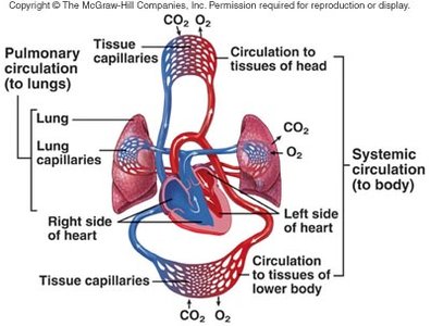
The heart is a muscular organ responsible for pumping blood throughout the body via two main circuits: pulmonary and systemic. It consists of four chambers: right atrium, right ventricle, left atrium, and left ventricle. - Right side: Receives deoxygenated blood and pumps it to the lungs (pulmonary circuit). - Left side: Receives oxygenated blood from the lungs and pumps it to the rest of the body (systemic circuit). Valves ensure unidirectional blood flow and prevent backflow.
Circulation of Blood Through the Heart
- Blood enters the right atrium via the superior and inferior vena cava. - Passes through the tricuspid valve to the right ventricle. - Pumped through the pulmonary valve into the pulmonary artery and to the lungs. - Oxygenated blood returns to the left atrium via pulmonary veins. - Passes through the bicuspid (mitral) valve to the left ventricle. - Pumped through the aortic valve into the aorta and systemic circulation. 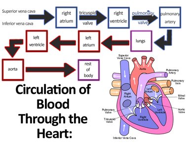
Cardiac Muscle Tissue
Characteristics of Cardiac Muscle Cells
Cardiac muscle cells are specialized for continuous rhythmic contraction. - Small, single central nucleus per cell. - Branching interconnections via intercalated discs. - Desmosomes: Prevent cell separation during contraction. - Gap junctions: Allow ions and action potentials to pass cell-to-cell, enabling the myocardium to contract as a unit. - Many mitochondria: Support aerobic metabolism. - Actin/myosin arrangement: Similar to skeletal muscle. - T-tubules: Wider and fewer than in skeletal muscle. - Smaller Ca2+ reserve than skeletal muscle. 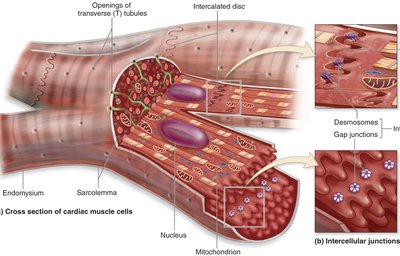
Cardiac Muscle Metabolism
Metabolic Adaptations
Cardiac muscle relies heavily on aerobic metabolism and must ensure adequate oxygen supply. - Myoglobin: Stores oxygen in muscle cells. - Coronary circulation: Supplies oxygenated blood to the heart muscle. - Adaptation to nutrients: Can utilize fatty acids, glucose, and even lactic acid for energy.
Cardiac Muscle Fibers
Types of Cardiac Fibers
There are two main types of cardiac muscle fibers: - Contractile fibers: Make up 99% of heart fibers; responsible for the heart's pumping activity. - Autorhythmic fibers: Depolarize spontaneously; control and coordinate heartbeat.
Action Potential in Cardiac Muscle
Phases of Action Potential in Contractile Cells
The action potential in ventricular contractile fibers consists of three phases: 1. Rapid depolarization: Voltage-gated fast Na+ channels open, Na+ enters, depolarization to +30 mV. 2. Plateau: Increased intracellular Ca2+ (from outside and SR), balances Na+ pumped out, maintains potential near 0 mV. 3. Repolarization: Ca2+ channels close, slow K+ channels open, K+ rushes out, repolarization occurs. 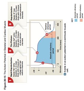
Contraction Mechanism
- Electrical activity (action potential) leads to mechanical contraction. - Two sources of Ca2+: Intracellular (SR) and extracellular.
Refractory Period
- Absolute refractory period: Membrane will not respond to a second stimulus. - Relative refractory period: Some response possible. - Long refractory period prevents tetanus, ensuring alternating contraction and relaxation. 
Autorhythmic Fibers and the Conducting System
Autorhythmic Fibers
- Make up 1% of cardiac cells. - Self-excitable: Set rhythm of heart (pacemaker function). - Part of the conduction system: Initiate and propagate impulses.
The Conducting System
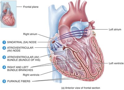
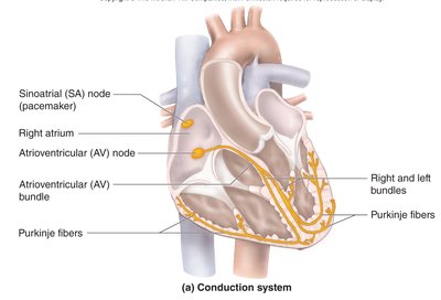
The heart's conducting system coordinates the heartbeat. - Sinoatrial (SA) node: Pacemaker, located in right atrium. - Atrioventricular (AV) node: Located in floor of right atrium. - AV bundle (bundle of His): Only pathway for action potential from atria to ventricles. - Bundle branches: Left and right, through interventricular septum. - Purkinje fibers: Spread impulse through ventricular myocardium. 
Sequence of Excitation
- SA node generates impulse. - Stimulus moves through internodal pathways to AV node. - Impulse pauses at AV node (allows atria to contract before ventricles). - AV bundle transmits impulse to ventricles. - Bundle branches and Purkinje fibers distribute impulse, causing ventricular contraction.
Action Potential in Nodal Rhythmic Fibers
Phases
1. Prepotential (pacemaker potential): Gradual depolarization toward threshold due to slow Na+ entry. 2. Fast depolarization: Voltage-gated Ca2+ channels open, Ca2+ enters. 3. Repolarization: Ca2+ channels close, K+ channels open, K+ exits.
Electrocardiogram (ECG/EKG)
ECG Waves and Heartbeat
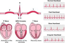
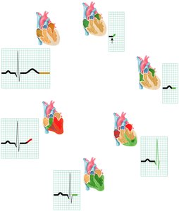
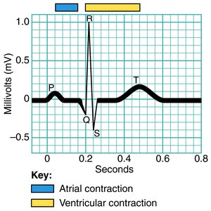
The ECG records all electrical events in the heart. - P wave: Atrial depolarization (contraction starts). - QRS complex: Ventricular depolarization (and atrial repolarization). - T wave: Ventricular repolarization. 
ECG Intervals
- P-Q (P-R) interval: Time from atrial excitation to ventricular excitation. - Q-T interval: Time from beginning of ventricular depolarization to end of repolarization.
Cardiac Arrhythmias
Types of Arrhythmias
- Bradycardia: Slow heart rate. - Tachycardia: Fast heart rate. - Fibrillation: Rapid, irregular contractions; poor pumping. - Premature Atrial Contractions (PACs): Early atrial contraction. - Paroxysmal Atrial Tachycardia (PAT): Flurry of atrial activity. - Atrial Fibrillation (AF): Atrial wall quivers, nonfunctional. - Premature Ventricular Contractions (PVCs): Early ventricular contraction. - Ventricular Tachycardia (VT): Multiple PVCs, serious. - Ventricular Fibrillation (VF): Cardiac arrest, fatal if untreated.
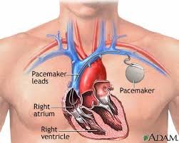
Artificial Pacemakers
- Used to correct abnormal heart rhythms. - Generate action potentials, bypass conduction system, couple atria and ventricles. 
The Cardiac Cycle
Phases of the Cardiac Cycle
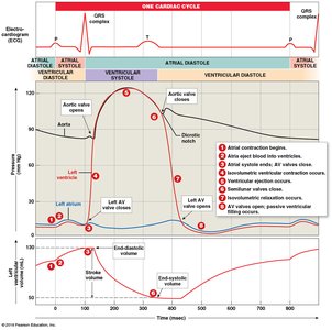
The cardiac cycle includes all events associated with one heartbeat (~800 msec at 75 bpm). - Systole: Contraction, blood ejected. - Diastole: Relaxation, chamber fills with blood. 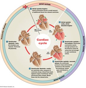
Heart Sounds
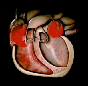
- S1 (lubb): AV valves close. - S2 (dubb): Semilunar valves close. - Heart murmur: Abnormal sound, indicates valve problems.
Volume Changes During Cardiac Cycle
- End-diastolic volume (EDV): Maximum blood in ventricles (~130 ml). - End-systolic volume (ESV): Blood remaining after contraction (~50 ml). - Stroke volume (SV): Blood ejected per beat.
Cardiac Output
Definition and Calculation
Cardiac output (CO) is the amount of blood pumped by each ventricle per minute. Example:
Factors Affecting Cardiac Output
- Heart rate (HR): Modified by autonomic nervous system and hormones. - Stroke volume (SV): Influenced by preload, contractility, and afterload.
Regulation of Stroke Volume
- Preload: Degree of stretch before contraction; increased by venous return and filling time. - Contractility: Forcefulness of contraction; increased by positive ionotropic agents (e.g., Ca2+, sympathetic stimulation). - Afterload: Pressure to overcome for ejection; increased by arterial resistance.
Frank-Starling Principle
- Increased EDV leads to increased SV: "More blood in = more blood out."
Effects of Aging and Heart Disease
Aging Effects
- Decreased cardiac output and heart rate. - Increased arrhythmias. - Hypertrophy of left ventricle. - Valve stenosis or incompetence. - Coronary artery disease and heart failure.
Congestive Heart Failure
- CO too low for adequate circulation. - Left-side failure: Pulmonary congestion (edema). - Right-side failure: Peripheral congestion (edema). - Fluid accumulation in either circuit.
Summary Table: Cardiac Cycle Phases
Phase | Event | Valve Status | Volume Change |
|---|---|---|---|
Atrial Systole | Atria contract, blood to ventricles | AV open, SL closed | Ventricles topped off (EDV) |
Ventricular Systole (early) | Ventricles contract, pressure rises | AV closed, SL closed | Isovolumetric contraction |
Ventricular Systole (late) | Blood ejected | AV closed, SL open | ESV reached |
Ventricular Diastole (early) | Ventricles relax | AV closed, SL closed | Isovolumetric relaxation |
Ventricular Diastole (late) | Ventricles fill passively | AV open, SL closed | 3/4 full by next cycle |
Additional info: The notes have been expanded with academic context, definitions, and examples to ensure completeness and clarity for college-level study.
