 Back
BackComprehensive Study Notes: The Reproductive System (ANP College Level)
Study Guide - Smart Notes
 Tailored notes based on your materials, expanded with key definitions, examples, and context.
The Reproductive System
Common Elements of the Male and Female Reproductive Systems
The reproductive system consists of primary reproductive organs (gonads) and accessory organs. Both sexes produce gametes and sex hormones, which are essential for sexual maturation and reproduction.
Gonads: Ovaries in females, testes in males; produce gametes (oocytes and sperm).
Sex Hormones: Influence maturation, development, and activity of reproductive organs.
Accessory Organs: Ducts transport gametes; in females, toward fertilization site; in males, toward the exterior.
Sexual Union: Copulation (coitus) allows gametes to unite for fertilization.
Female Reproductive Tract: Provides support, protection, and nourishment if fertilization occurs.
Sexual Maturation and Puberty
Puberty marks the onset of sexual maturity, triggered by hormonal changes. The hypothalamus secretes GnRH, stimulating the anterior pituitary to release FSH and LH, which in turn activate the gonads.
FSH and LH: Initiate gamete and sex hormone production.
Gamete Production: Females release one oocyte monthly; males produce millions of sperm daily.
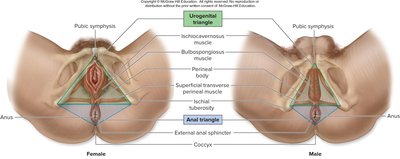
Anatomy of the Perineum
The perineum is a diamond-shaped region between the thighs, divided into two triangles: the urogenital triangle and the anal triangle. It contains muscles and structures relevant to both sexes.
Boundaries: Pubic symphysis (anterior), ischial tuberosities (lateral), coccyx (posterior).
Urogenital Triangle: Contains external genitalia and associated muscles.
Anal Triangle: Contains the anus and external anal sphincter.
Gametogenesis and Heredity
Gametogenesis
Gametogenesis is the process of forming human sex cells (gametes) via meiosis. Females produce secondary oocytes, while males produce sperm.
Meiosis: Cell division that reduces chromosome number by half, producing haploid gametes.
Heredity and Chromosomes
Hereditary information is carried on 23 pairs of chromosomes: 22 pairs of autosomes and 1 pair of sex chromosomes (XX for females, XY for males).
Diploid Cells (2n): 23 pairs of chromosomes.
Haploid Cells (n): 23 single chromosomes; gametes are haploid.
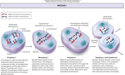
Meiosis I: Reduction Division
Meiosis I separates homologous chromosomes, resulting in two cells with 23 chromosomes each (still paired as sister chromatids).
Prophase I: Homologous chromosomes pair and undergo crossing over, increasing genetic diversity.
Metaphase I: Tetrads align at the cell midline; random assortment occurs.
Anaphase I: Homologous chromosomes are pulled to opposite poles.
Telophase I and Cytokinesis: Nuclear envelopes reform, and the cell divides.
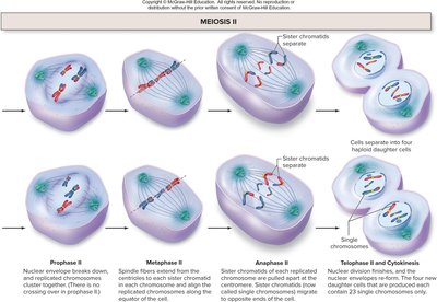
Meiosis II: Separation of Sister Chromatids
Meiosis II resembles mitosis, separating sister chromatids to produce four haploid cells.
Prophase II: Chromosomes condense; no crossing over.
Metaphase II: Chromosomes align at the midline.
Anaphase II: Sister chromatids are pulled apart.
Telophase II and Cytokinesis: Four haploid cells are formed.
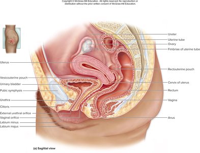
Female Reproductive System
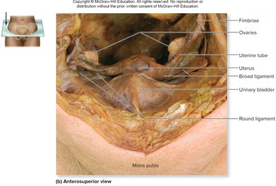
Overview of Female Pelvic Anatomy
The female pelvis contains primary and accessory reproductive organs, including the ovaries, uterine tubes, uterus, vagina, and external genitalia.
Vesicouterine Pouch: Space between bladder and uterus.
Rectouterine Pouch: Space between rectum and uterus.
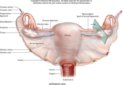
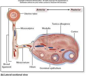
Ovaries: Structure and Function
Ovaries are paired organs responsible for oocyte production and sex hormone release. They are anchored by several ligaments and supplied by ovarian arteries and veins.
Mesovarium: Attaches ovary to broad ligament.
Ovarian Ligament: Anchors ovary to uterus.
Suspensory Ligament: Attaches ovary to pelvic wall.
Germinal Epithelium: Outer layer; tunica albuginea is the dense CT capsule beneath.
Cortex: Contains ovarian follicles.
Medulla: Contains blood vessels, lymphatics, and nerves.
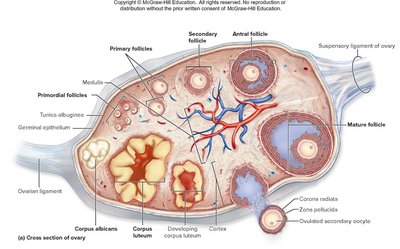
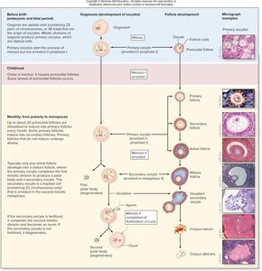
Ovarian Follicles: Types and Development
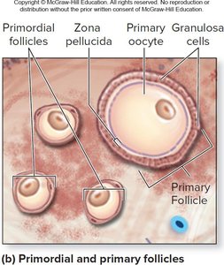
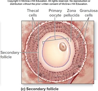
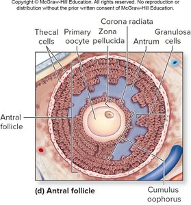
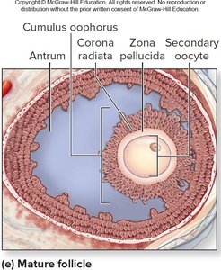
Ovarian follicles are structures containing oocytes at various stages of development, surrounded by supporting cells.
Primordial Follicle: Primary oocyte with a single layer of flattened cells; most primitive type.
Primary Follicle: Primary oocyte with cuboidal granulosa cells; secretes estrogen.
Secondary Follicle: Primary oocyte with multiple granulosa layers and thecal cells.
Antral Follicle: Primary oocyte, granulosa cells, and fluid-filled antrum; surrounded by zona pellucida and corona radiata.
Mature Follicle: Contains secondary oocyte; completes meiosis I and arrests in metaphase II.
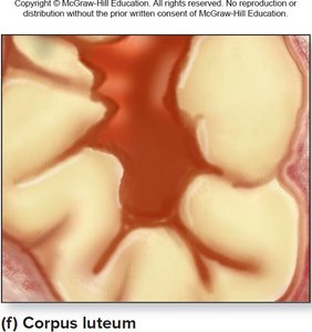
Corpus Luteum: Formed after ovulation; secretes progesterone and estrogen.
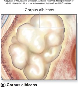
Corpus Albicans: Degenerated corpus luteum; connective tissue scar.
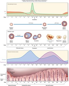
Oogenesis and the Ovarian Cycle
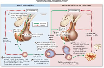
Oogenesis is the maturation of a primary oocyte to a secondary oocyte, occurring in stages throughout a female's life. The ovarian cycle consists of three phases: follicular, ovulation, and luteal.
Follicular Phase: Days 1–13; follicles mature under FSH and LH stimulation.
Ovulation: Day 14; release of secondary oocyte induced by LH surge.
Luteal Phase: Days 15–28; corpus luteum secretes hormones, prepares uterus for implantation.
Menopause: Cessation of ovarian cycles, typically between ages 45–55.
Hormonal Regulation of the Ovarian Cycle
The ovarian cycle is regulated by a feedback loop involving the hypothalamus, pituitary gland, and ovaries. GnRH stimulates FSH and LH release, which promote follicle maturation and hormone secretion.
Estrogen: Assists follicle development; positive feedback increases LH for ovulation.
Progesterone: Secreted by corpus luteum; maintains uterine lining.
Inhibin: Inhibits FSH to prevent excessive follicle development.
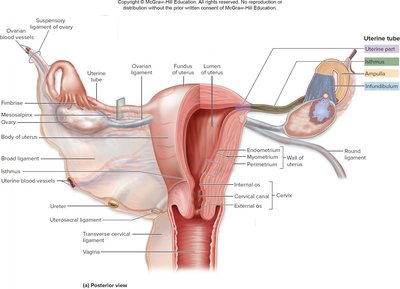
Uterine Tubes, Uterus, and Vagina
The uterine tubes transport oocytes, the uterus supports embryo development, and the vagina serves as the birth canal and site of intercourse.
Uterine Tubes: Infundibulum, ampulla (site of fertilization), isthmus, and uterine part.
Uterus: Fundus, body, isthmus, cervix; three tunics (perimetrium, myometrium, endometrium).
Vagina: Fibromuscular tube; mucosa, muscularis, adventitia; acidic secretions prevent infection.
Uterine (Menstrual) Cycle and Menstruation
The uterine cycle involves cyclical changes in the endometrial lining, influenced by estrogen and progesterone.
Menstrual Phase: Days 1–5; functional layer sloughs off.
Proliferative Phase: Days 6–14; new functional layer develops.
Secretory Phase: Days 15–28; increased progesterone, uterine gland development.

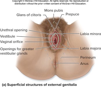
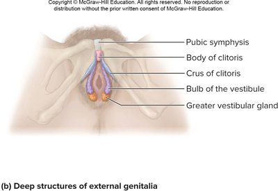
External Genitalia
The external genitalia (vulva) include the mons pubis, labia majora, labia minora, vestibule, clitoris, and associated glands.
Mons Pubis: Fatty area anterior to pubic symphysis.
Labia Majora: Thick folds homologous to male scrotum.
Labia Minora: Thin, vascular folds internal to labia majora.
Clitoris: Erectile body with sensory receptors.
Vestibular Glands: Secrete mucus for lubrication.
Mammary Glands
Mammary glands are tubuloalveolar exocrine glands that produce breast milk for infant nutrition. They are composed of lobes, lobules, alveoli, and ducts.
Nipple: Central projection with duct openings.
Areola: Pigmented ring around nipple.
Suspensory Ligaments: Support breast structure.
Lactation: Prolactin stimulates milk production; oxytocin triggers milk ejection.
*Additional info: Mammary gland structure and function are essential for postnatal nutrition and are regulated by hormonal changes during pregnancy and after birth.*
Male Reproductive System
Scrotum and Testes
The scrotum is a skin-covered sac providing a cooler environment for sperm development. The testes produce sperm and androgens, and are covered by tunica vaginalis and tunica albuginea.
Spermatic Cord: Contains blood vessels, nerves, and three layers of fascia.
Pampiniform Plexus: Veins that cool arterial blood.
Temperature Regulation: Dartos and cremaster muscles adjust testis position.
*Additional info: Proper temperature is critical for spermatogenesis; scrotal muscles respond to environmental changes to maintain optimal conditions.*
Testes and Spermatogenesis
Spermatogenesis is the process of sperm development within seminiferous tubules, regulated by hormones.
Sustentacular Cells: Support sperm development; release inhibin.
Interstitial Cells: Produce testosterone under LH stimulation.
Blood-Testis Barrier: Protects developing sperm from immune response.
*Additional info: Spermatogenesis begins at puberty and continues throughout life, producing millions of sperm daily.*
Hormonal Regulation of Spermatogenesis
GnRH from the hypothalamus stimulates FSH and LH release, which promote spermatogenesis and androgen production.
FSH: Stimulates sustentacular cells to produce ABP.
LH: Stimulates interstitial cells to produce testosterone.
Testosterone: Facilitates spermatogenesis, secondary sex characteristics, and libido.
Inhibin: Released by sustentacular cells; inhibits FSH.
*Additional info: Negative feedback mechanisms regulate hormone levels to maintain reproductive function.*
Spermatogenesis and Spermiogenesis
Spermatogenesis involves mitosis and meiosis to produce four haploid sperm from each diploid spermatogonium. Spermiogenesis is the maturation of spermatids into spermatozoa.
Spermatogonia: Diploid cells divide by mitosis.
Primary Spermatocytes: Undergo meiosis I.
Secondary Spermatocytes: Undergo meiosis II.
Spermatids: Mature into spermatozoa.
*Additional info: Female gametogenesis produces one viable oocyte; male gametogenesis produces four sperm per cycle.*
Duct System and Accessory Glands
The male duct system transports sperm from the testes to the urethra, with accessory glands producing seminal fluid.
Rete Testis, Efferent Ductules, Epididymis: Sperm maturation and storage.
Ductus Deferens: Transports sperm; forms ampulla.
Ejaculatory Duct: Conducts sperm and seminal fluid to urethra.
Urethra: Prostatic, membranous, and spongy regions.
Seminal Vesicles: Secrete alkaline fluid with fructose and prostaglandins.
Prostate Gland: Secretes citric acid, seminalplasmin, PSA.
Bulbourethral Glands: Produce mucus for lubrication.
*Additional info: Semen is a combination of sperm and seminal fluid; 200–500 million sperm are released per ejaculation.*
Penis Anatomy and Male Sexual Response
The penis contains three erectile bodies and is involved in sexual intercourse and urination. Sexual response includes excitement, orgasm, and resolution phases.
Corpora Cavernosa: Paired dorsal erectile bodies.
Corpus Spongiosum: Ventral body containing urethra.
Glans: Tip of penis; contains external urethral orifice.
Excitement Phase: Erection via increased blood flow.
Orgasm: Ejaculation of semen.
Resolution: Relaxation and refractory period.
*Additional info: Male sexual response is regulated by parasympathetic and sympathetic nervous systems.*
Genetic and Phenotypic Sex Determination
Genetic vs. Phenotypic Sex
Genetic sex is determined by sex chromosomes (XX or XY) at fertilization. Phenotypic sex refers to the appearance of internal and external genitalia, influenced by the SRY gene on the Y chromosome.
SRY Gene: Initiates male development by stimulating androgen production.
Absence of SRY: Results in female development.
*Additional info: Sex determination occurs during embryonic development, with differentiation of gonads and genitalia.*
Internal and External Genitalia Development
Development of internal and external genitalia is guided by genetic and hormonal factors during embryogenesis.
Female Development: Paramesonephric ducts form uterus and vagina; mesonephric ducts degenerate.
Male Development: SRY gene induces testis formation; anti-Müllerian hormone inhibits paramesonephric ducts; mesonephric ducts form male duct system.
Gubernaculum: Assists testis descent into scrotum.
*Additional info: External genitalia differentiate by week 12–20 of development.*
Puberty, Menopause, and Male Climacteric
Puberty is the period when reproductive organs become fully functional. Menopause marks the end of female reproductive cycles, while male climacteric is a gradual decline in testosterone and reproductive function.
Puberty: Initiated by GnRH, FSH, and LH; external sex characteristics develop.
Menopause: Cessation of menstruation; decreased estrogen and progesterone.
Male Climacteric: Gradual decline in testosterone; may cause mood changes and decreased libido.
*Additional info: Hormonal changes during puberty and aging affect reproductive health and function.* ----------------------------------------
