 Back
BackComprehensive Study Notes: The Respiratory System (Anatomy & Physiology)
Study Guide - Smart Notes
 Tailored notes based on your materials, expanded with key definitions, examples, and context.
The Respiratory System
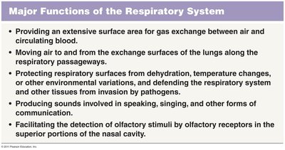
Major Functions of the Respiratory System
The respiratory system is essential for gas exchange, sound production, and protection of respiratory surfaces. It also plays a role in olfaction and defending the body from environmental hazards.
Gas Exchange: Provides an extensive surface area for the exchange of oxygen and carbon dioxide between air and circulating blood.
Ventilation: Moves air to and from the exchange surfaces of the lungs along the respiratory passageways.
Protection: Protects respiratory surfaces from dehydration, temperature changes, and invasion by pathogens.
Sound Production: Produces sounds involved in speaking, singing, and other forms of communication.
Olfaction: Facilitates the detection of olfactory stimuli by olfactory receptors in the superior portions of the nasal cavity.
Functional Anatomy of the Respiratory System
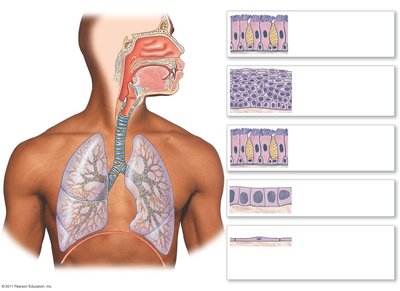
The respiratory system is divided into anatomical and functional regions, each with specialized structures and roles.
Conducting Portion: Extends from the nasal cavity to the bronchioles; responsible for filtering, warming, and humidifying air.
Respiratory Portion: Includes the smallest bronchioles and alveoli, where gas exchange occurs.
Respiratory Defense System and Epithelia
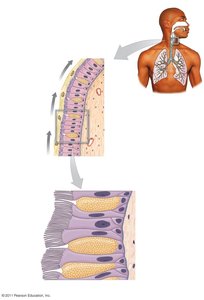
The respiratory tract is lined with specialized epithelia and defense mechanisms to protect delicate gas exchange surfaces.
Pseudostratified Ciliated Epithelium: Lines much of the conducting portion; cilia move mucus and trapped debris toward the pharynx (the 'mucus escalator').
Mucous Cells: Secrete mucus to trap particles larger than ~5 µm.
Lamina Propria: Connective tissue layer supporting the epithelium.
Stratified Squamous Epithelium: Found in the inferior pharynx, protecting against abrasion.
Cuboidal Epithelium: Present in finer bronchioles.
Simple Squamous Epithelium: Lines alveoli, facilitating rapid gas exchange.
Upper Respiratory System
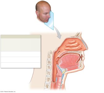
The upper respiratory system filters, warms, and humidifies incoming air, protecting the lower respiratory tract.
Nose: Primary passageway for air; contains nasal cartilages and external nares (nostrils).
Nasal Cavity: Contains an extensive network of veins to warm air and mucus to humidify it. Connects to paranasal sinuses, which help moisturize and clean surfaces.
Nasal Septum: Divides the nasal cavity into left and right portions (formed by the perpendicular plate of the ethmoid and vomer).
Meatuses: Passageways between nasal conchae that swirl incoming air, trapping particles (superior, middle, inferior).
Pharynx: Shared chamber for respiratory and digestive systems; divided into nasopharynx, oropharynx, and laryngopharynx.
Other Structures: Hard and soft palate, glottis, larynx, and trachea.
Larynx
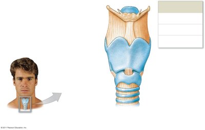
The larynx protects the glottis and is involved in sound production. It is composed of several cartilages and ligaments.
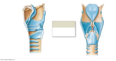
Large Unpaired Cartilages: Epiglottis (prevents entry of solids/liquids), thyroid cartilage (Adam’s apple), cricoid cartilage (provides support and muscle attachment).
Small Paired Cartilages: Cuneiform, corniculate, and arytenoid cartilages (support vocal and vestibular folds).
Vocal Folds: Contain vocal ligaments for sound production; vestibular folds prevent foreign objects from entering the glottis.
Speech Production: Involves phonation (larynx), articulation (tongue, teeth, lips), and resonance (pharynx, oral/nasal cavities, sinuses).
Trachea and Bronchi
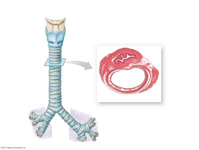
The trachea is a flexible tube supported by C-shaped cartilages, branching into primary bronchi that enter the lungs.
Tracheal Rings: Prevent collapse and overexpansion; connected by trachealis muscle for flexibility during swallowing.
Primary Bronchi: Lead to each lung and branch into secondary and tertiary bronchi, eventually forming bronchioles.
Bronchioles: Lack cartilage, have thick smooth muscle layers, and allow for bronchoconstriction/dilation.
Lung Gross Anatomy
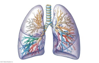
The lungs are divided into lobes and bronchopulmonary segments, each supplied by tertiary bronchi.
Left Lung: Two lobes (superior, inferior) separated by the oblique fissure.
Right Lung: Three lobes (superior, middle, inferior) separated by horizontal and oblique fissures.
Hilum: Medial groove for passage of bronchi, vessels, nerves, and lymphatics.
Pulmonary Lobules and Alveoli
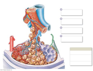
Pulmonary lobules are the smallest functional units of the lung, containing alveoli where gas exchange occurs.
Terminal Bronchioles: Supply each lobule and branch into respiratory bronchioles.
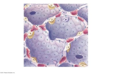
Alveoli: Surrounded by capillaries and elastic fibers; main site of gas exchange.
Alveolar Structure: Type I pneumocytes (simple squamous for gas exchange), Type II pneumocytes (secrete surfactant to reduce surface tension), and alveolar macrophages (defense).
Respiratory Membrane: Consists of alveolar epithelium, fused basal laminae, and capillary endothelium; very thin to facilitate diffusion.
Pleura
The pleura are serous membranes that reduce friction and allow smooth lung movement during breathing.
Visceral Pleura: Covers the outer surface of the lungs.
Parietal Pleura: Lines the inner surface of the thoracic cavity.
Pleural Fluid: Lubricates and reduces friction between pleural surfaces.
Respiratory Physiology: Ventilation and Gas Exchange
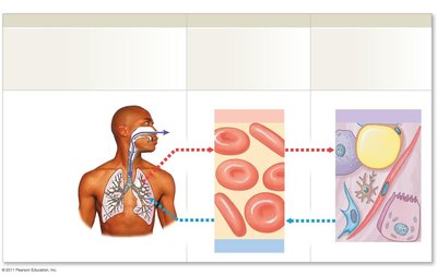
Respiration involves external and internal processes for gas exchange and transport.
External Respiration: Exchange of gases between interstitial fluids and the external environment (includes pulmonary and alveolar ventilation).
Internal Respiration: Oxygen absorption and carbon dioxide production by tissue cells.
Gas Diffusion: Occurs across the respiratory membrane and capillary walls.
Pulmonary Ventilation: Mechanics and Pressures
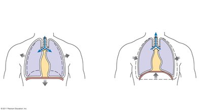
Ventilation is driven by pressure changes resulting from volume changes in the thoracic cavity, described by Boyle’s law.
Boyle’s Law: (Pressure is inversely proportional to volume at constant temperature).
Inhalation: Diaphragm contracts, thoracic volume increases, pressure decreases, air flows in.
Exhalation: Diaphragm relaxes, thoracic volume decreases, pressure increases, air flows out.
Pleural Fluid: Maintains lung adhesion to thoracic wall; loss leads to atelectasis (lung collapse).
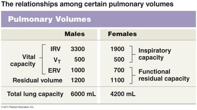
Respiratory Volumes and Capacities
Respiratory volumes and capacities describe the amount of air moved during different phases of the respiratory cycle.
Tidal Volume (VT): Air moved in/out during resting breathing (~500 mL).
Inspiratory Reserve Volume (IRV): Additional air inhaled after a normal inspiration.
Expiratory Reserve Volume (ERV): Additional air exhaled after a normal expiration.
Residual Volume: Air remaining after maximal exhalation.
Vital Capacity:
Total Lung Capacity:
Gas Laws and Partial Pressures
Gas movement is governed by several physical laws:
Dalton’s Law: Each gas in a mixture exerts its own partial pressure; total pressure is the sum of all partial pressures.
Henry’s Law: The amount of gas dissolved in a liquid is proportional to its partial pressure.
Oxygen and Carbon Dioxide Transport
Oxygen and carbon dioxide are transported in the blood by different mechanisms.
Oxygen: 98.5% bound to hemoglobin (Hb), forming oxyhemoglobin (HbO2); 1.5% dissolved in plasma.
Oxygen-Hemoglobin Saturation Curve: S-shaped curve showing % saturation at different PO2 values; affected by pH (Bohr effect), temperature, and BPG.
Carbon Dioxide: 7% dissolved in plasma, 23% bound to Hb (carbaminohemoglobin), 70% as bicarbonate ion (HCO3-) via carbonic anhydrase reaction.
Key Equation:
Respiratory Control Mechanisms
Breathing is regulated by neural centers and reflexes at multiple levels:
Medulla Oblongata: Respiratory rhythmicity centers (DRG and VRG) control basic rhythm and accessory muscles.
Pons: Apneustic and pneumotaxic centers modulate depth and rate of breathing.
Higher Centers: Hypothalamus, limbic system, and cortex can override lower centers.
Reflexes: Chemoreceptors (pH, PCO2, PO2), baroreceptors (blood pressure), stretch receptors, and protective reflexes (coughing, sneezing).
Clinical Considerations: Pulmonary Disease and Aging
Diseases and aging can significantly affect respiratory function.
Compliance: Measure of lung expandability; reduced by diseases affecting lung tissue or chest wall.
Resistance: Force required to move air; increased by bronchoconstriction, mucus, or inflammation.
COPD: Includes asthma (bronchoconstriction, mucus), chronic bronchitis (inflammation, mucus), and emphysema (alveolar destruction).
Aging: Decreases elasticity, vital capacity, and increases risk of emphysema.
Smoking: Major risk factor for lung cancer and emphysema; damages cilia, increases mucus, and leads to irreversible tissue changes.
Table: Pulmonary Volumes (Males vs. Females)
Pulmonary Volumes | Males | Females |
|---|---|---|
Inspiratory Reserve Volume (IRV) | 3300 mL | 1900 mL |
Tidal Volume (VT) | 500 mL | 500 mL |
Expiratory Reserve Volume (ERV) | 1000 mL | 700 mL |
Residual Volume | 1200 mL | 1100 mL |
Total Lung Capacity | 6000 mL | 4200 mL |
Additional info: This guide covers the anatomical and physiological foundations of the respiratory system, including clinical relevance and regulatory mechanisms, suitable for ANP college-level study.
