 Back
Conduction System of the Heart and Electrocardiogram (ECG/EKG): Study Notes
Study Guide - Smart Notes
 Tailored notes based on your materials, expanded with key definitions, examples, and context.
Conduction System of the Heart
Overview of the Cardiac Conduction System
The conduction system of the heart is responsible for initiating and distributing electrical impulses that regulate the heart's rhythm and coordinate contraction. This system is composed of specialized autorhythmic muscle cells that ensure the heart beats in a coordinated and efficient manner.
Autorhythmicity: The ability of certain cardiac muscle cells to spontaneously generate action potentials without external stimulation.
Purpose: To maintain a consistent heart rate and synchronize the contraction of cardiac muscle tissue.
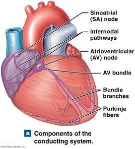
Main Components of the Conduction System
Sinoatrial (SA) Node: Located in the right atrium; acts as the natural pacemaker, establishing the basic heart rhythm (sinus rhythm).
Atrioventricular (AV) Node: Receives impulses from the SA node and delays them slightly to allow atrial contraction before ventricular contraction.
AV Bundle (Bundle of His): Conducts impulses from the AV node to the bundle branches.
Right and Left Bundle Branches: Carry impulses down the interventricular septum toward the apex of the heart.
Purkinje Fibers: Distribute the electrical impulse throughout the ventricles, causing coordinated ventricular contraction.
Electrocardiogram (ECG/EKG)
Principles and Methodology of ECG
An electrocardiogram (ECG or EKG) is a diagnostic tool that records the electrical activity of the heart over time. It is used to assess heart rhythm, detect abnormalities, and monitor cardiac health.
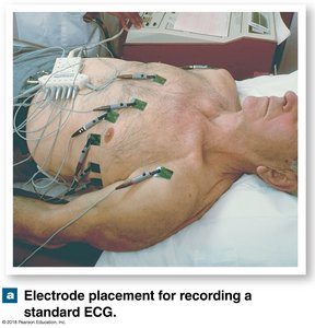
Electrodes: Placed at specific locations on the body surface to detect electrical signals generated by the heart.
Leads: Different combinations of electrode placements provide multiple views (leads) of the heart's electrical activity.
Clinical Use: Abnormal ECG patterns can indicate arrhythmias, myocardial infarction, and other cardiac disorders.
Electrode Placement for Standard ECG
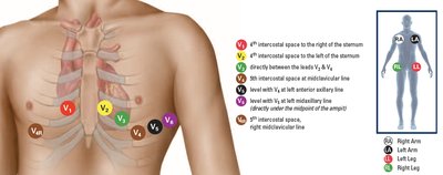
Proper electrode placement is essential for accurate ECG recordings. A standard 12-lead ECG uses 10 electrodes: 4 limb electrodes and 6 precordial (chest) electrodes.
Limb Electrodes: Placed on the right arm (RA), left arm (LA), right leg (RL), and left leg (LL).
Precordial Electrodes (V1–V6): Placed at specific intercostal spaces on the chest to monitor the heart from different angles.
ECG Waves and Intervals
Components of the ECG
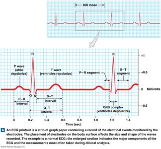
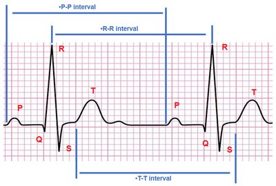
The ECG tracing consists of several characteristic waves and intervals, each corresponding to specific electrical and mechanical events in the cardiac cycle.
Wave/Complex | Electrical Event | Mechanical Event |
|---|---|---|
P wave | Atrial depolarization | Atrial systole (contraction) |
QRS complex | Ventricular depolarization (atrial repolarization is masked) | Ventricular systole (contraction), atrial diastole (relaxation) |
T wave | Ventricular repolarization | Ventricular diastole (relaxation) |
Key Intervals and Segments
P-P Interval: Time between consecutive P waves; reflects atrial rhythm.
R-R Interval: Time between consecutive R waves; used to calculate heart rate.
T-T Interval: Time between consecutive T waves; reflects ventricular rhythm.
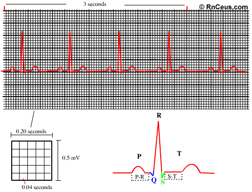
P-R Interval: Time from onset of atrial depolarization to onset of ventricular depolarization.
S-T Segment: Time between ventricular depolarization and repolarization.
Calculating Cardiac Cycle Duration and Heart Rate
Standard ECG Measurements
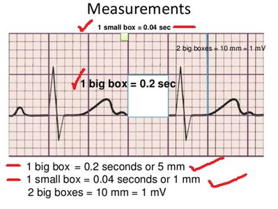
ECG paper is standardized so that each small box represents 0.04 seconds (1 mm), and each large box represents 0.2 seconds (5 mm). These measurements are used to calculate the duration of cardiac cycles and heart rate.
Duration of one cardiac cycle: Multiply the number of millimeters (mm) between two R waves (R-R interval) by 0.04 seconds.
Heart rate calculation: Divide 60 seconds by the duration of one cardiac cycle (in seconds).
Formulas:
Duration of one cardiac cycle:
Heart rate:

Example Calculation
Given: R-R interval = 17.5 mm
Duration of one cardiac cycle: s
Heart rate: beats/min (within normal range)
Normal and Abnormal Heart Rates
Classification of Heart Rates
Normal heart rate: 60–100 beats/min
Tachycardia: Heart rate > 100 beats/min
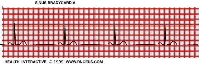
Bradycardia: Heart rate < 60 beats/min
Practice Questions and Answers
Identify the structural components of the conducting system of the heart:
A. SA node
B. AV node
C. AV bundle (Bundle of His)
D. Right & Left bundle branch
E. Purkinje fibers
Identify the wave at the pointer:
A. P wave
B. QRS complex
C. T wave
Calculate the duration of one cardiac cycle and the heart rate on the EKG running at the standard speed:
Q3: A. 0.65 s, B. 0.70 s, C. 0.72 s, D. 0.80 s
Q4: A. 82.5 beats/min, B. 83.3 beats/min, C. 85.7 beats/min, D. 87.5 beats/min
Sample Calculation:
R-R interval = 18 mm
Duration of one cardiac cycle = s
Heart rate = beats/min (normal range)
Additional info: The conduction system and ECG interpretation are foundational for understanding cardiac physiology and diagnosing arrhythmias in clinical practice.
