 Back
Connective Tissue: Structure, Classification, and Histology
Study Guide - Smart Notes
 Tailored notes based on your materials, expanded with key definitions, examples, and context.
Connective Tissue Overview
Definition and General Structure
Connective tissue is one of the four primary tissue types in the human body, providing structural support, protection, and integration of various tissues and organs. It is characterized by the presence of cells, protein fibers, and ground substance, collectively forming the extracellular matrix.
Cells: Includes fibroblasts, adipocytes, macrophages, and mesenchymal cells.
Protein fibers: Reticular, collagen, and elastic fibers provide strength and flexibility.
Ground substance: Amorphous material that fills the space between cells and fibers; contains blood vessels.
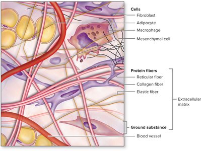
Components of Connective Tissue
Fibroblasts: Synthesize fibers and ground substance.
Adipocytes: Store fat and provide insulation.
Macrophages: Phagocytic cells involved in immune defense.
Mesenchymal cells: Undifferentiated cells that can develop into other cell types.
Collagen fibers: Provide tensile strength.
Elastic fibers: Allow tissues to stretch and recoil.
Reticular fibers: Form supportive networks in soft tissues.
Embryonic Connective Tissue
Mesenchyme
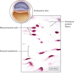
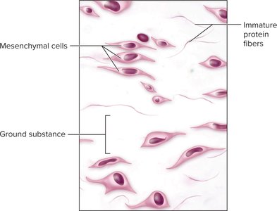
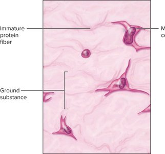
Mesenchyme is the first connective tissue to appear in the embryo, consisting of mesenchymal cells and immature protein fibers within a ground substance. It serves as the origin for all other connective tissues.
Mesenchymal cells: Star-shaped, multipotent cells.
Immature protein fibers: Not fully developed, provide initial structural support.
Ground substance: Gel-like matrix.
Mucous Connective Tissue
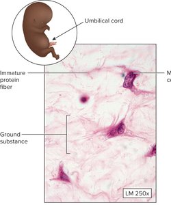
Mucous connective tissue is found in the umbilical cord and is similar to mesenchyme but with more abundant ground substance.
Location: Umbilical cord.
Cells: Mesenchymal cells.
Fibers: Immature protein fibers.
Ground substance: More abundant and gelatinous.
Classification of Connective Tissue
Major Types
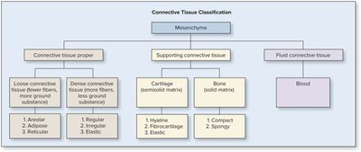
Connective tissue is classified into three main categories: connective tissue proper, supporting connective tissue, and fluid connective tissue.
Connective tissue proper: Includes loose and dense connective tissues.
Supporting connective tissue: Includes cartilage and bone.
Fluid connective tissue: Includes blood.
Connective Tissue Proper
Loose connective tissue: Areolar, adipose, and reticular types; more ground substance, fewer fibers.
Dense connective tissue: Regular, irregular, and elastic types; more fibers, less ground substance.
Supporting Connective Tissue
Cartilage: Hyaline, fibrocartilage, and elastic types; semisolid matrix.
Bone: Compact and spongy types; solid matrix.
Fluid Connective Tissue
Blood: Contains cells suspended in plasma.
Loose Connective Tissue
Areolar Tissue
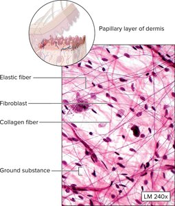
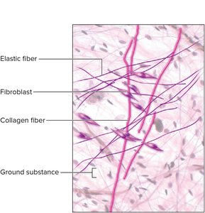
Areolar tissue is a common type of loose connective tissue found beneath epithelial layers, providing support and nourishment.
Cells: Fibroblasts, macrophages, adipocytes.
Fibers: Collagen, elastic, and reticular.
Ground substance: Gel-like matrix.
Adipose Tissue
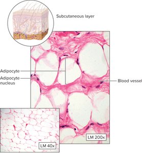
Adipose tissue is specialized for fat storage and insulation, found in the subcutaneous layer.
Cells: Adipocytes (fat cells).
Function: Energy storage, insulation, and protection.
Blood vessels: Present to supply nutrients.
Dense Connective Tissue
Dense Regular Connective Tissue
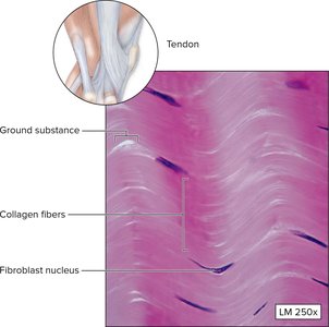
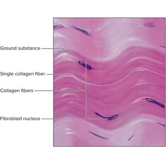
Dense regular connective tissue is composed of parallel collagen fibers, providing tensile strength in one direction. It is found in tendons and ligaments.
Collagen fibers: Arranged in parallel bundles.
Fibroblast nuclei: Located between fibers.
Ground substance: Minimal.
Dense Irregular Connective Tissue
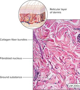
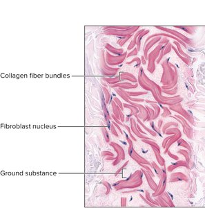
Dense irregular connective tissue contains collagen fibers arranged in multiple directions, providing strength in various directions. It is found in the reticular layer of the dermis.
Collagen fiber bundles: Irregular arrangement.
Fibroblast nuclei: Scattered throughout.
Ground substance: Present but less abundant.
Supporting Connective Tissue
Cartilage
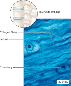
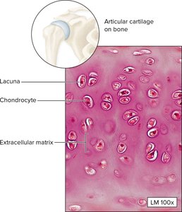
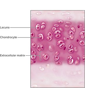
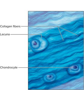
Cartilage is a flexible, semisolid supporting tissue found in joints, intervertebral discs, and other structures.
Cells: Chondrocytes located in lacunae.
Matrix: Extracellular matrix with collagen fibers.
Types: Hyaline cartilage (articular surfaces), fibrocartilage (intervertebral discs), elastic cartilage (ear).

Bone
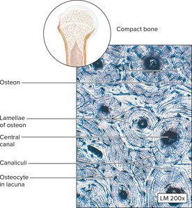
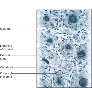
Bone is a rigid supporting tissue with a solid matrix, providing structural support and protection.
Cells: Osteocytes located in lacunae.
Matrix: Mineralized extracellular matrix.
Structure: Osteons, lamellae, central canal, canaliculi.
Fluid Connective Tissue
Blood
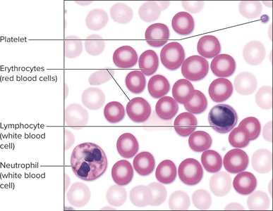
Blood is a fluid connective tissue composed of cells suspended in plasma, responsible for transport of gases, nutrients, and waste.
Cells: Erythrocytes (red blood cells), leukocytes (white blood cells), platelets.
Plasma: Liquid matrix.
Functions: Transport, immune defense, clotting.

Muscular Tissue (Brief Overview)
Skeletal Muscle
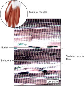
Skeletal muscle tissue is composed of long, cylindrical fibers with multiple nuclei and striations, responsible for voluntary movement.
Fibers: Striated appearance due to arrangement of actin and myosin.
Nuclei: Multiple, peripherally located.
Cardiac Muscle
Cardiac muscle tissue is found in the heart wall, composed of branched cells with intercalated discs, striations, and centrally located nuclei.
Intercalated discs: Specialized junctions for synchronized contraction.
Striations: Present.
Nuclei: Usually one per cell, centrally located.
Summary Table: Connective Tissue Classification
Type | Subtypes | Main Features |
|---|---|---|
Connective Tissue Proper | Loose (Areolar, Adipose, Reticular) Dense (Regular, Irregular, Elastic) | Fibers and ground substance; support and binding |
Supporting Connective Tissue | Cartilage (Hyaline, Fibrocartilage, Elastic) Bone (Compact, Spongy) | Semisolid or solid matrix; structural support |
Fluid Connective Tissue | Blood | Cells in liquid matrix; transport |
Key Terms and Concepts
Extracellular matrix: Combination of fibers and ground substance outside cells.
Lacuna: Small cavity housing a cell (chondrocyte or osteocyte).
Osteon: Structural unit of compact bone.
Chondrocyte: Cartilage cell.
Fibroblast: Cell that produces fibers and ground substance.
Additional info:
Connective tissue is essential for tissue repair, immune response, and metabolic functions.
Histological identification relies on cell type, fiber arrangement, and matrix composition.
