 Back
BackConnective Tissues: Structure, Function, and Types
Study Guide - Smart Notes
 Tailored notes based on your materials, expanded with key definitions, examples, and context.
Connective Tissue Overview
Introduction to Connective Tissue
Connective tissue (CT) is one of the four primary tissue types in the human body. It is highly abundant, found in every organ, and exhibits a wide variety of forms and functions. The main roles of connective tissue include supporting, connecting, and separating different types of tissues and organs in the body.
Abundance: Present in all organs and tissues.
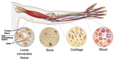
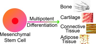
Diversity: Includes bone, cartilage, blood, adipose, and more.
Functions: Connects tissues, forms the skeleton, stores and transports nutrients, supports blood vessels and nerves.
Functions of Connective Tissue
Major Functions
Connective tissues perform a variety of essential functions in the body, depending on their specific type and location.
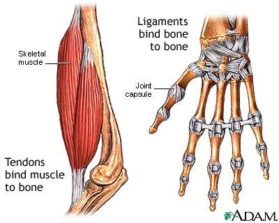
Connection: Ligaments connect bone to bone; tendons connect muscle to bone.
Support: Bone and cartilage provide structural support for the body.
Transport: Blood transports nutrients, gases, and waste products.
Storage: Adipose tissue stores energy; bone stores minerals.
Protection: Adipose cushions organs; bone protects vital structures.
Defense: Loose areolar CT is involved in immune responses and inflammation.
Structural Elements of Connective Tissue
Cells and Extracellular Matrix
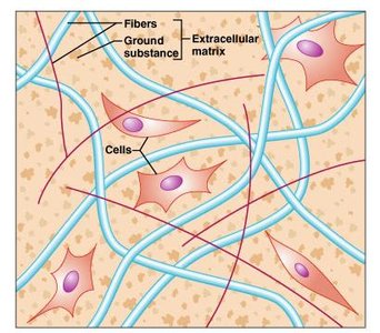
All connective tissues are composed of two main components: cells and an abundant extracellular matrix (ECM). The ECM is the non-living component that separates the living cells and determines the tissue's properties.
Cells: The living component; type varies by tissue (e.g., fibroblasts, chondrocytes, osteocytes, adipocytes).
Extracellular Matrix: Composed of ground substance and protein fibers; provides structural and biochemical support.
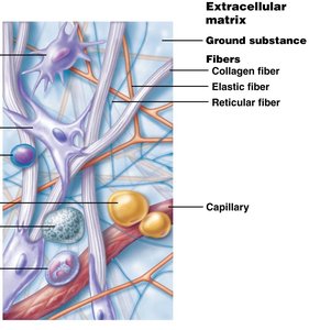
Ground Substance and Protein Fibers
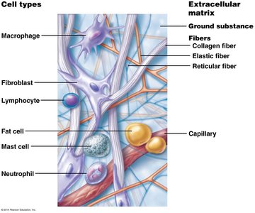
The extracellular matrix consists of ground substance and three main types of protein fibers, each contributing to the tissue's function and mechanical properties.
Ground Substance: Amorphous material that fills the space between cells and fibers; can be jelly-like (CT proper, cartilage), fluid (blood), or mineralized (bone).
Protein Fibers:
Collagen fibers: Strongest and most abundant; provide tensile strength.
Elastic fibers: Allow stretch and recoil.
Reticular fibers: Short, thin fibers forming supportive networks.
Origin and Classification of Connective Tissue
Embryonic Origin
All connective tissues originate from a common embryonic tissue called mesenchyme. Mesenchymal stem cells are multipotent and can differentiate into various connective tissue types, including bone, cartilage, adipose, and others.
Classification of Connective Tissues
Connective tissues are classified based on their structure and function into four main categories:
Connective tissue proper: Loose and dense types
Cartilage: Hyaline, fibrocartilage, elastic
Bone (Osseous tissue): Compact and spongy
Blood
Cells of Connective Tissue
Major Cell Types and Functions
Each connective tissue type has a primary cell responsible for producing and maintaining the extracellular matrix. The cell names reflect their function:
"-blast": Producing the ECM (e.g., fibroblast, chondroblast, osteoblast)
"-cyte": Maintaining the ECM (e.g., fibrocyte, chondrocyte, osteocyte)
"-clast": Breaking down the ECM (e.g., osteoclast, chondroclast)
Other important cells include defense cells (macrophages, lymphocytes), fat cells (adipocytes), and blood cells.
Types of Connective Tissue Proper
Loose Connective Tissue
Loose connective tissue is characterized by a loose arrangement of fibers and abundant ground substance. It includes areolar, adipose, and reticular tissues.
Areolar: Most widespread; supports and binds other tissues, holds body fluids, defends against infection.
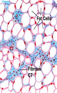
Adipose: Composed mostly of adipocytes; stores energy, insulates, and cushions organs.
Reticular: Contains only reticular fibers; forms a soft internal skeleton (stroma) in lymphoid organs.
Dense Connective Tissue
Dense connective tissue contains more fibers and less ground substance, providing greater strength and resistance to stretching. It is subdivided into:
Dense regular CT: Collagen fibers run parallel; found in tendons and ligaments; resists tension in one direction.
Dense irregular CT: Collagen fibers run in multiple directions; found in dermis, joint capsules; resists tension from various directions.
Dense elastic CT: Dominated by elastic fibers; found in elastic arteries; allows stretch and recoil.
Specialized Connective Tissues
Cartilage
Cartilage is a firm, flexible tissue with a matrix that is mostly water, allowing it to spring back after compression. The main cell types are chondroblasts, chondrocytes, and chondroclasts. Types include hyaline, fibrocartilage, and elastic cartilage.
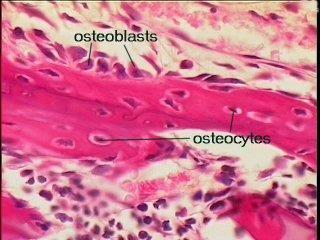
Bone (Osseous Tissue)
Bone tissue has a mineralized matrix composed of calcium salts and collagen fibers, making it solid yet flexible. The main cell types are osteoblasts, osteocytes, and osteoclasts.
Blood
Blood is considered a connective tissue because it develops from mesenchyme and consists of blood cells surrounded by a nonliving matrix (plasma). Blood cells are produced in red bone marrow.
Clinical Application: Fibrodysplasia Ossificans Progressiva (FOP)
Definition and Pathology
Fibrodysplasia Ossificans Progressiva (FOP) is a rare genetic disorder in which fibrous tissues such as ligaments, tendons, and muscles are progressively replaced by bone, leading to immobility and other complications. The name literally means "abnormal growth of bone formation that progresses."
Fibro-: Fiber
Dysplasia: Abnormal growth
Ossificans: Bone formation
Progressiva: Progressing
With FOP, normal connective tissues are replaced by bone, severely restricting movement and leading to early mortality.
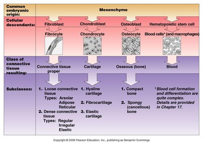
Summary Table: Connective Tissue Types and Origins
Common Embryonic Origin | Cellular Descendants | Class of Connective Tissue Resulting | Subclasses |
|---|---|---|---|
Mesenchyme | Fibroblast → Fibrocyte | Connective tissue proper | Loose (areolar, adipose, reticular), Dense (regular, irregular, elastic) |
Mesenchyme | Chondroblast → Chondrocyte | Cartilage | Hyaline, fibrocartilage, elastic |
Mesenchyme | Osteoblast → Osteocyte | Osseous (bone) | Compact, spongy |
Mesenchyme | Hematopoietic stem cell → Blood cells (and macrophages) | Blood | --- |
