 Back
BackDefense Mechanisms of the Immune System: Innate and Adaptive Immunity
Study Guide - Smart Notes
 Tailored notes based on your materials, expanded with key definitions, examples, and context.
Defense Mechanisms of the Immune System
Introduction to Body Defenses
The human body is constantly exposed to a variety of pathogens and foreign substances. To protect itself, the body employs a complex system of defense mechanisms, which are broadly categorized into innate (non-specific) and adaptive (specific) immunity. These mechanisms work together to prevent entry, detect, and eliminate harmful agents.
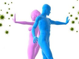
Innate (Non-Specific) Immunity
Physical and Chemical Barriers
Physical and chemical barriers are the body's first line of defense, preventing the entry of pathogens.
Skin: Acts as a physical barrier, preventing pathogen entry.
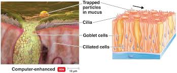
Mucous membranes: Trap and remove microbes from the respiratory and digestive tracts.
Ciliary escalator: Cilia in the upper respiratory tract move mucus and trapped particles toward the mouth for expulsion.
Tears and sweat: Contain enzymes like lysozyme that destroy bacteria.
Stomach acid (HCl): Destroys ingested pathogens.
Normal flora: Compete with pathogens for resources and space.
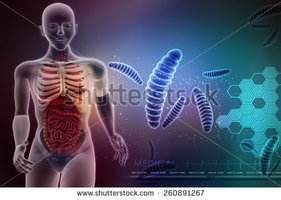
Lymphatic System and Lymphatic Vessels
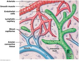
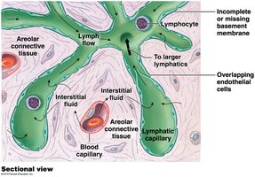
The lymphatic system is essential for fluid balance and immune defense. Lymphatic vessels collect interstitial fluid (lymph) and return it to the bloodstream, filtering it through lymph nodes where immune responses can be initiated.
Lymphatic capillaries: Closed at one end, have larger diameters and thinner walls than blood capillaries, and allow one-way entry of fluid, solutes, and even pathogens.
One-way valves: Prevent backflow and ensure lymph moves toward the venous system.
Cellular Defenses
Several types of white blood cells provide non-specific cellular defense:
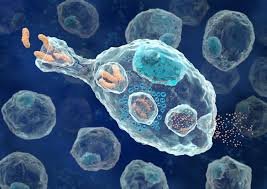
Macrophages and neutrophils: Phagocytic cells that engulf and destroy pathogens and debris. They are the first line of cellular defense.
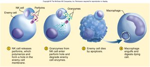
Natural Killer (NK) cells: Lymphocytes that destroy infected or abnormal cells by releasing cytotoxic chemicals, but are not phagocytic.
Inflammatory Response
Inflammation is a localized, non-specific response to tissue damage or infection. Its main functions are to dilute harmful substances, bring oxygen and nutrients for tissue repair, and allow immune cells to access the affected area.
Cardinal signs: Redness, warmth, swelling, pain.
Chemical mediators: Histamine, prostaglandins, and kinins increase blood flow and vessel permeability.

Fever
Fever is an elevation of body temperature above 37.2°C (99°F), induced by pyrogens. It can inhibit pathogen growth and accelerate tissue repair and immune responses.

Chemical Defenses: Complement System and Interferons
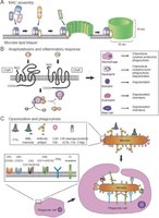
Complement system: A group of over 30 proteins that enhance immune responses by promoting cell lysis, inflammation, and phagocytosis (opsonization).
Interferons: Proteins released by virus-infected cells that inhibit viral replication and activate NK cells and macrophages.
Adaptive (Specific) Immunity
General Properties
Adaptive immunity is characterized by specificity, versatility, memory, and tolerance. It targets specific antigens and provides long-lasting protection.
Specificity: Each lymphocyte recognizes only one antigen.
Memory: Memory cells enable a faster, stronger response upon re-exposure to the same antigen.
Tolerance: Immune system ignores self-antigens but attacks non-self antigens.
Origin of Lymphocytes
All lymphocytes originate from stem cells in the bone marrow. They differentiate into:
B cells: Mature in bone marrow; responsible for humoral immunity (antibody production).
T cells: Mature in the thymus; responsible for cell-mediated immunity.
Steps of the Immune Response
Antigen challenge: Lymphocyte recognizes and binds to an antigen.
Cell activation: Lymphocyte is activated and undergoes clonal selection (rapid division).
Differentiation: Cloned cells become effector cells (attack invaders) or memory cells (for future responses).
T Cells: Cell-Mediated Immunity
T cells attack infected or abnormal cells directly. Types of T cells include:
Helper T cells: Secrete chemicals to enhance immune response and activate B cells.
Cytotoxic (Killer) T cells: Destroy infected cells by releasing cytotoxic chemicals.
Memory T cells: Provide long-term immunity by remembering antigens.
Suppressor (Regulator) T cells: Turn off immune response to prevent autoimmunity.
B Cells: Humoral Immunity
B cells differentiate into plasma cells that produce antibodies and memory cells. Antibodies circulate in body fluids and bind to specific antigens, marking them for destruction.
Antibody (immunoglobulin) classes:
IgG: Most abundant; crosses placenta.
IgE: Involved in allergies and inflammation.
IgD: Functions as a B cell receptor.
IgM: First antibody produced after antigen exposure.
IgA: Found in secretions (mucus, saliva, breast milk).
Active vs. Passive Immunity
Type | Source of Antibodies | Memory Cells Formed? | Duration |
|---|---|---|---|
Active Immunity | Produced by the individual's own immune system | Yes | Long-lasting |
Passive Immunity | Received from another source (e.g., mother, injection) | No | Temporary |
Immune System Disorders
Transplant Rejection
Transplanted tissues may be rejected if recognized as non-self. Matching tissues and immunosuppressive drugs can increase acceptance. The thymus gland plays a role in establishing self-tolerance during fetal development.
Allergies (Hypersensitivity)
Immediate hypersensitivity: Mediated by IgE, causes rapid allergic reactions (e.g., hay fever, anaphylaxis).
Delayed hypersensitivity: Involves T cells, appears 1-3 days after exposure (e.g., contact dermatitis).
Immunodeficiency Diseases
SCID (Severe Combined Immunodeficiency Disease): Lack of functional T and B cells.
HIV/AIDS: Destroys helper T cells, leading to severe immunodeficiency.
Autoimmune Diseases
Multiple sclerosis: Immune system attacks myelin in CNS.
Myasthenia gravis: Antibodies attack acetylcholine receptors.
Rheumatoid arthritis: Chronic inflammation of joints.
Type I Diabetes: Immune system destroys insulin-producing pancreatic cells.
Repair and Healing
Scar tissue: Collagen fibers replace damaged tissue but lack original function.
Pus: Accumulation of dead neutrophils and tissue debris.
Abscess: Walled-off pocket of pus.
Granuloma: Walled-off area containing indigestible substances.
