 Back
BackDevelopment and Inheritance
Study Guide - Smart Notes
 Tailored notes based on your materials, expanded with key definitions, examples, and context.
Development and Inheritance
Fertilization and Early Embryonic Development
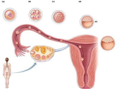
Fertilization is the process by which the genetic material from a sperm merges with that of an ovum, resulting in the formation of a zygote. This event occurs in the fallopian (uterine) tube within 12 to 24 hours after ovulation. The penetration of the sperm triggers a release of calcium ions (Ca2+), which induces changes in the oocyte wall (zona pellucida) to prevent further sperm entry. The secondary oocyte completes Meiosis II to become an ovum, and the fertilized ovum is referred to as the zygote.
Zona Pellucida: Protective glycoprotein layer surrounding the oocyte.
Zygote: The initial one-cell structure formed after fertilization.
Cleavage: Mitotic divisions of the zygote without an increase in size, leading to the morula stage.
Morula and Blastocyst Stages
Following fertilization, the zygote undergoes cleavage to form a morula, a solid ball of cells by day 4. By day 5, a fluid-filled cavity develops within the morula, forming the blastocyst. Differentiation occurs, resulting in two main cell types:
Trophoblast: Becomes the chorion, part of the embryonic membranes, and secretes human Chorionic Gonadotropin (hCG) to maintain the corpus luteum.
Inner Cell Mass: Develops into the embryo.
Implantation: The blastocyst implants into the endometrium around day 6.

Gastrulation and Germ Layer Formation
By day 15, the inner cell mass differentiates into three primary germ layers through the process of gastrulation:
Ectoderm: Forms the epidermis and nervous system.
Mesoderm: Develops into muscle, bone, connective tissue, and peritoneum.
Endoderm: Forms the lining (mucosa) of the respiratory and gastrointestinal tracts, and other organs.
Organogenesis and neurulation (formation of the brain and spinal cord from ectoderm) begin during this stage.
Embryonic Membranes and Placenta Development
Several embryonic membranes form to support the developing embryo:
Amnion: Produces amniotic fluid, serving as a shock absorber.
Chorionic Villi: Fetal part of the placenta, secretes proteins to block maternal antibody production and hCG.
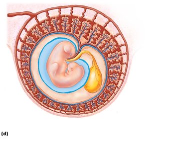
Yolk Sac: Provides nutrients and blood cells in early development.
The placenta develops as chorionic villi containing blood vessels grow into the endometrium. Maternal and fetal blood remain separate but close enough for diffusion. The umbilical cord contains two arteries (carrying deoxygenated blood and wastes away from the fetus) and one vein (carrying oxygen and nutrients to the fetus).
Fetal Circulation
Fetal circulation is uniquely adapted to prenatal life, with several vascular modifications that are occluded at birth:
Ductus Venosus: Bypasses the liver, allowing umbilical vein blood to drain into the inferior vena cava.
Foramen Ovale: Opening in the interatrial septum, bypasses pulmonary circulation.
Ductus Arteriosus: Bypasses pulmonary circulation, connecting the pulmonary trunk to the aorta.
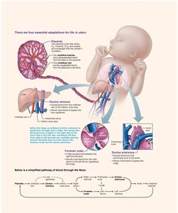
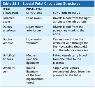
Table: Special Fetal Circulation Structures
Fetal Structure | Postnatal Structure | Function in Fetus |
|---|---|---|
Foramen ovale | Fossa ovalis | Diverts blood from the right atrium to the left atrium |
Ductus arteriosus | Ligamentum arteriosum | Diverts blood from the pulmonary trunk to the aorta |
Ductus venosus | Ligamentum venosum | Carries blood from the umbilical vein through the liver (bypassing sinusoids) into the inferior vena cava |
Umbilical arteries | Median umbilical ligaments | Paired vessels carry blood from the fetus to the placenta |
Umbilical vein | Round ligament of the liver (ligamentum teres) | Single vessel carries oxygenated blood from the placenta to the fetus |
Placental and Maternal Hormones
Hormones play a crucial role in pregnancy and fetal development:
Human Chorionic Gonadotropin (hCG): Secreted by the chorion, maintains corpus luteum and stimulates progesterone and estrogen production.
Relaxin: Increases flexibility of joints and dilates cervix during labor.
Estrogen/Progesterone: Maintain endometrium, prepare mammary glands for lactation, and keep cervix closed.
Prolactin: Stimulates milk production, inhibited during pregnancy by progesterone.
Oxytocin: Stimulates milk release and myometrial contraction, maintained by positive feedback.
Clinical Considerations: Preeclampsia
Preeclampsia is a dangerous complication of pregnancy characterized by hypertension, proteinuria, and peripheral edema. It can progress to eclampsia, which involves generalized seizures and can be fatal. The only cure is delivery of the baby.
Inheritance and Genetic Principles
Mendelian Genetics
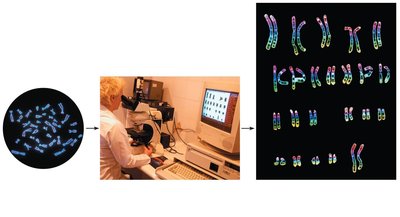
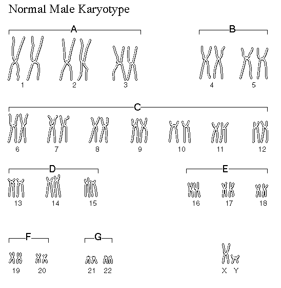
Mendelian genetics describes inheritance patterns where one gene codes for one trait. All somatic cells contain 23 pairs of chromosomes, including autosomes (body traits) and sex chromosomes (XX for females, XY for males). Each chromosome contains many genes, and alternative forms of genes are called alleles.
Allele: Alternative form of a gene.
Homozygous: Paired alleles are identical.
Heterozygous: Paired alleles are different.
Dominant: Allele always expressed when present (capital letter).
Recessive: Allele only expressed in homozygous condition.
Phenotype: Physical expression of a trait.
Genotype: Actual alleles present on a chromosome.
Autosomal Recessive and Dominant Inheritance
Autosomal recessive inheritance requires two recessive alleles for the trait to be expressed (e.g., PKU: pp). Autosomal dominant inheritance requires only one dominant allele for the trait to be expressed (e.g., Huntington's Chorea: HH or Hh).
Non-Mendelian Inheritance Patterns
Incomplete Dominance: Neither allele is dominant; phenotype is a blend (e.g., Sickle Cell Anemia: Ss = mild form).
Co-Dominance: Both alleles are equally expressed (e.g., Blood type AB).
Sex-Linked: Genes appear only on the X chromosome (e.g., color blindness, hemophilia).
Nondisjunction and Aneuploidy
Nondisjunction is the failure of chromosome pairs to separate during meiosis, resulting in aneuploidy (added or deleted chromosomes). Examples include:
Trisomy 21 (Down's Syndrome): Extra chromosome 21.
Turner's Syndrome: XO (missing sex chromosome).
Klinefelter's Syndrome: XXY (extra sex chromosome).
Polygenic Inheritance
Polygenic inheritance involves traits controlled by the combined effects of many genes, such as skin, eye, and hair color, height, and body build.
Additional info: Academic context was added to clarify the stages of embryonic development, genetic terminology, and inheritance patterns.
