 Back
BackDigestive System Part 2: Stomach, Liver, Gallbladder, Pancreas, and Small Intestine
Study Guide - Smart Notes
 Tailored notes based on your materials, expanded with key definitions, examples, and context.
The Stomach
Structure and Function
The stomach is a muscular, hollow organ that serves as a temporary storage tank for food and initiates the chemical breakdown of proteins. It produces up to 3 liters of acidic gastric juice per day, which contains hydrochloric acid (HCl), pepsin, and, in infants, rennin.
HCl and Pepsin: Begin the denaturation and chemical digestion of proteins.
Gastric Lipase: Accounts for up to 15% of lipid digestion.
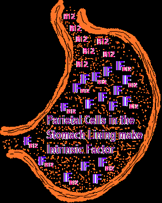
Intrinsic Factor: Essential for vitamin B12 absorption; deficiency leads to pernicious anemia.
Chyme Formation: Muscular mixing and digestive action convert food into chyme.
Absorption: Limited to alcohol and aspirin, which are lipid-soluble.
Regulation of Gastric Secretion
Gastric secretion is regulated by neural and hormonal mechanisms, involving both long (vagus nerve) and short reflexes, as well as hormones such as gastrin and histamine.
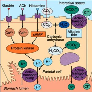
Parietal Cells: Express receptors for gastrin, acetylcholine (ACh), and histamine; histamine is the key stimulator of acid secretion.
Proton Pump: Target for acid secretion on the apical surface of parietal cells.
Phases of Gastric Secretion: Cephalic (preparatory), gastric (most active), and intestinal (winding down).
Cephalic Phase
Triggered by aroma, taste, sight, or thought of food before it enters the stomach.
Gastric Phase
Lasts 3–4 hours, provides two-thirds of gastric juice.
ACh and gastrin stimulate histamine secretion.
Very low gastric pH (<2) or increased sympathetic activity reduces acid secretion.
Intestinal Phase
Begins with brief stimulation (intestinal gastrin) followed by inhibition.
Inhibitory factors: duodenal distension, presence of acidic, fatty, or hypertonic chyme.
Inhibition via enterogastric reflex and enterogastrones (secretin, CCK).
Mechanism of HCl Secretion
Parietal cells secrete HCl through a complex mechanism involving carbonic anhydrase, proton pumps, and chloride channels.
Carbonic Anhydrase: Catalyzes the formation of carbonic acid from CO2 and H2O.
H+-K+ ATPase: Pumps H+ into the stomach lumen in exchange for K+.
Alkaline Tide: HCO3- leaves the cell, causing a temporary increase in blood pH.
Cl- Channels: Allow chloride ions to enter the lumen and combine with H+ to form HCl.
Secretion of Pepsin by Chief Cells
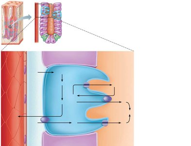
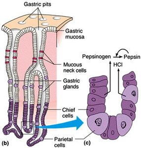
Chief cells secrete pepsinogen, an inactive precursor stored in zymogen granules. Once secreted, pepsinogen is activated to pepsin in the acidic environment of the stomach.
Pepsin: Hydrolyzes proteins to peptides; optimal pH is 2–3.
Primary Stimulus: Acetylcholine (ACh).
Regulation of Gastric Motility and Emptying
The stomach stretches to accommodate incoming food, with pressure remaining constant until about 1.5 liters are ingested.
Receptive Relaxation: Reflex-mediated relaxation coordinated by the swallowing center in the brainstem.
Gastric Accommodation: Intrinsic ability of smooth muscle to exhibit stress-relaxation response.
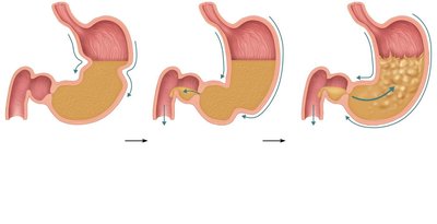
Peristaltic Waves: Move toward the pylorus at a rate of 3 per minute, set by enteric pacemaker cells.
Chyme Delivery: Only liquids and small particles pass through the pyloric valve; most chyme is forced backward into the stomach.
Regulation of Gastric Emptying
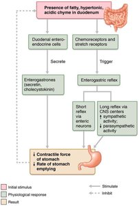
The duodenum controls the rate of gastric emptying to prevent overfilling.
Duodenal Receptors: Respond to stretch and chemical signals.
Enterogastric Reflex and Enterogastrones: Inhibit gastric secretion and duodenal filling.
Emptying Time: Stomach empties in about 4 hours; fatty chyme increases this time.
Carbohydrate-rich Chyme: Moves quickly through the duodenum.
Liver, Gallbladder, and Pancreas
Liver
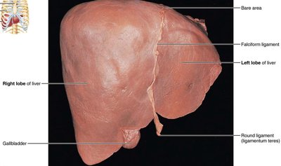
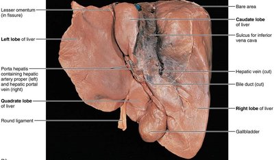
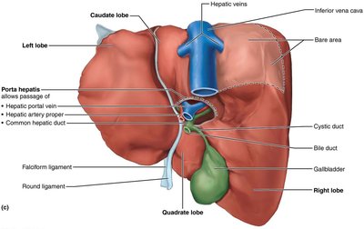
The liver is the largest gland in the body (~1.4 kg) and occupies most of the right hypochondriac and epigastric regions.
Lobes: Right, left, caudate, quadrate.
Falciform Ligament: Separates left and right lobes and suspends the liver from the diaphragm and anterior body wall.
Visceral Peritoneum: Encloses the liver except for the bare area.
Bile Production: Liver produces about 900 ml of bile daily.
Composition of Bile
Bile is a yellow-green, alkaline solution containing bile salts, bile pigments, cholesterol, neutral fats, phospholipids, and electrolytes.
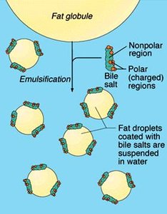
Bile Salts and Phospholipids: Aid in lipid digestion; main bile salts are cholic acid and chenodeoxycholic acid (cholesterol derivatives).
Amphipathic Molecules: Bile salts have both polar and nonpolar regions, allowing them to emulsify fats.
Bile Pigment: Main pigment is bilirubin, converted to urobilinogen or stercobilin by intestinal bacteria.
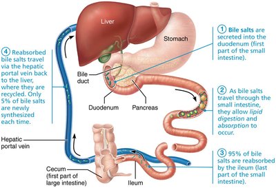
Bile Salt Recycling: About 95% of bile salts are recycled via enterohepatic circulation.
Gallbladder
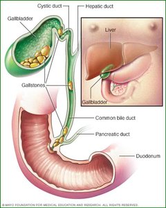
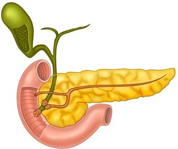
The gallbladder is a thin-walled muscular sac that stores and concentrates bile by absorbing water and ions.
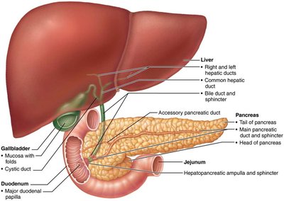
Bile Flow: Muscular contractions push bile into the cystic duct, then into the bile duct to enter the duodenum via the hepatopancreatic sphincter.
Gallstones: Supersaturation of bile salts with cholesterol, combined with stasis, can lead to cholesterol crystallization and formation of gallstones (biliary calculi).
Pancreas
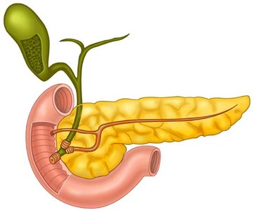
The pancreas is mostly retroperitoneal, with its head encircled by the duodenum and its tail abutting the spleen.
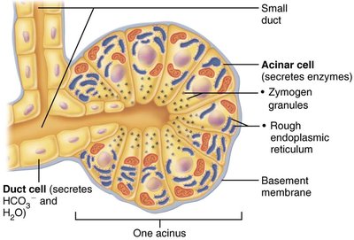
Pancreatic Juice: Produces 1200–1500 ml daily; contains enzymes and bicarbonate.
Acinar Cells: Produce digestive enzymes (zymogen granules).
Duct Cells: Secrete bicarbonate to neutralize stomach acid.
Activation of Pancreatic Proteases
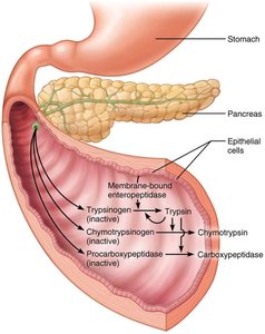
Proteases are secreted in inactive forms and activated in the duodenum by enteropeptidase, a brush border enzyme.
Trypsinogen: Activated to trypsin, which then activates chymotrypsinogen and procarboxypeptidase.
Other Enzymes: Amylase, lipases, nucleases.
Regulation of Bile and Pancreatic Secretion
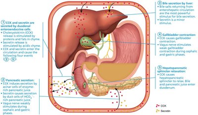
Hepatopancreatic Sphincter: Closed when no digestion occurs; bile backs up into the gallbladder.
Cholecystokinin (CCK): Responds to fatty chyme in the duodenum, stimulating gallbladder contraction, pancreatic juice secretion, and relaxation of the sphincter.
Secretin: Stimulates secretion of bicarbonate-rich fluid from epithelial cells lining bile and pancreatic ducts.
Bile Salt Recycling: Major stimulus for bile production and secretion by the liver.
The Small Intestine
Structure and Function
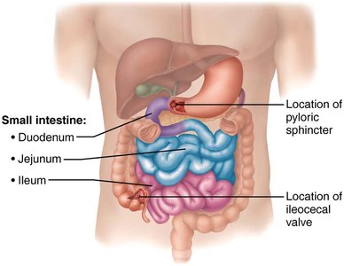
The small intestine is the primary site for digestion and absorption, extending from the pyloric sphincter to the ileocecal valve.
Length: 6–7 meters (in situ, 2–4 meters due to muscle tone).
Diameter: 2.5–4 cm.
Regions: Duodenum (~25 cm), jejunum (~2.5 m), ileum (~3.6 m).
Blood Supply: Superior mesenteric artery; veins drain into hepatic portal vein.
Modifications for Absorption
The small intestine has structural modifications to increase surface area for absorption:
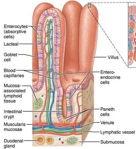
Circular Folds: Permanent folds of mucosa and submucosa; slow down food movement.
Villi: Finger-like projections; longest in the duodenum.
Microvilli: Cytoplasmic extensions forming the brush border; contain important enzymes.
Histology of Small Intestine Wall
Intestinal Crypts: Tubular glands scattered between villi.
Cell Types:
Enterocytes: Absorptive cells with tight junctions and microvilli; absorb breakdown products of digestion.
Goblet Cells: Produce mucus.
Enteroendocrine Cells: Source of enterogastrones (secretin, CCK).
Paneth Cells: Secrete antimicrobial agents (defensins, lysozyme).
Stem Cells: Continuously divide to replace all cell types; villus epithelium renewed every 2–4 days.
MALT: Mucosa-associated lymphoid tissue protects against microorganisms; includes lymphoid follicles and Peyer's patches.
Lamina Propria: Contains plasma cells that secrete IgA.
Submucosa: Areolar connective tissue; duodenal glands secrete alkaline mucus.
Muscularis Externa: Typical two layers.
Intestinal Juice: 1–2 liters produced daily; slightly alkaline (pH 7.4–7.8), mostly water, electrolytes, and mucus.
Major Stimulus: Hyperosmotic and/or acidic chyme.
Summary Table: Key Digestive Functions
Organ | Main Function | Key Secretions |
|---|---|---|
Stomach | Protein digestion, chyme formation | HCl, pepsin, intrinsic factor |
Liver | Bile production | Bile salts, bilirubin |
Gallbladder | Bile storage/concentration | None (stores bile) |
Pancreas | Enzyme and bicarbonate secretion | Proteases, amylase, lipase, nucleases, HCO3- |
Small Intestine | Absorption, enzyme secretion | Intestinal juice, brush border enzymes |
Additional info: The notes have been expanded to provide academic context, definitions, and examples for clarity and completeness.
