 Back
BackDigestive System Part 2: The Stomach, Liver, Gallbladder, Pancreas, and Small Intestine
Study Guide - Smart Notes
 Tailored notes based on your materials, expanded with key definitions, examples, and context.
The Stomach
Structure and Function
The stomach serves as a temporary storage tank and produces up to 3 liters of acidic gastric juice per day. It initiates the chemical breakdown of proteins through the secretion of HCl and pepsin. In infants, rennin is also secreted. The stomach's muscular action and digestive secretions convert food into a semi-liquid substance called chyme. Although the stomach does not absorb many substances, alcohol and aspirin are exceptions due to their lipid solubility.
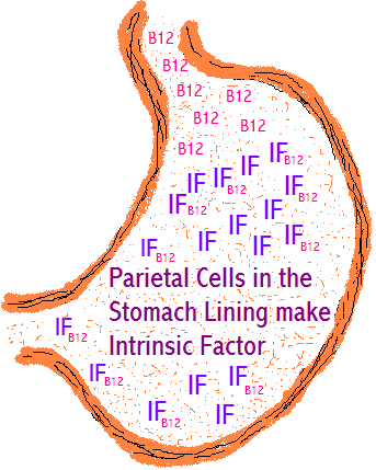
Intrinsic Factor (IF): Secreted by parietal cells, intrinsic factor is essential for vitamin B12 absorption in the small intestine. Without it, pernicious anemia develops, making this the only gastric function essential to life.
Regulation of Gastric Secretion
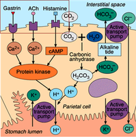
Gastric secretion is regulated by neural and hormonal mechanisms. Both long (vagus nerve-mediated) and short reflexes are involved, with hormonal control primarily via gastrin and histamine. Parietal cells have receptors for three stimulatory chemicals: acetylcholine (ACh), gastrin, and histamine. Histamine is the most potent stimulator, targeting the proton pump on the apical surface of parietal cells.
Three Phases of Gastric Secretion:
Cephalic Phase: Triggered by the sight, smell, taste, or thought of food; prepares the stomach for incoming food.
Gastric Phase: Begins when food enters the stomach; accounts for two-thirds of gastric juice secretion. Both ACh and gastrin stimulate histamine release, enhancing acid secretion.
Intestinal Phase: Starts as chyme enters the duodenum. Initially stimulates, then inhibits gastric secretion to prevent overloading the intestine.
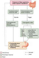
Inhibition of Gastric Secretion: Achieved by the enterogastric reflex and enterogastrones (e.g., secretin, CCK) in response to duodenal distension, acidic, fatty, or hypertonic chyme.
Mechanism of HCl Secretion by Parietal Cells
Parietal cells secrete HCl through a process involving the enzyme carbonic anhydrase, which catalyzes the formation of carbonic acid from CO2 and H2O. The dissociation of carbonic acid yields H+ and HCO3-. H+ is pumped into the stomach lumen via the H+-K+ ATPase (proton pump), while HCO3- is exchanged for Cl- in the interstitial fluid, resulting in the "alkaline tide" in blood.
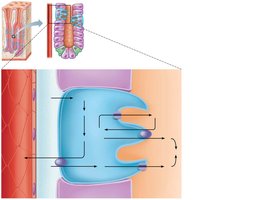
Equation: $\mathrm{CO_2 + H_2O \xrightarrow{carbonic\ anhydrase} H_2CO_3 \rightarrow H^+ + HCO_3^-}$
Secretion of Pepsin by Chief Cells
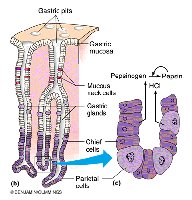
Pepsinogen is secreted by chief cells as an inactive precursor and is activated to pepsin in the acidic environment of the stomach. Pepsin hydrolyzes proteins into peptides, with an optimal pH of 2–3. Acetylcholine is the primary stimulus for pepsin secretion.
Regulation of Gastric Motility and Emptying
The stomach stretches to accommodate food, with pressure remaining constant up to 1.5 L due to receptive relaxation and gastric accommodation. Peristaltic waves, set by the Basic Electrical Rhythm (BER) from pacemaker cells, move chyme toward the pylorus. Only small amounts of chyme pass into the duodenum at a time; the rest is retropulsed for further mixing.
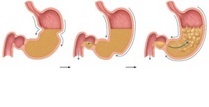
Duodenal Control: The duodenum regulates gastric emptying by responding to stretch and chemical signals, inhibiting gastric secretion and motility via the enterogastric reflex and enterogastrones.
Gastric Emptying Time: Typically 4 hours, but fatty chyme can delay emptying to 6 hours or more. Carbohydrate-rich chyme empties more quickly.
Liver, Gallbladder, and Pancreas
Overview and Functions
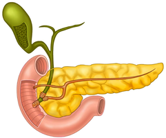
The liver, gallbladder, and pancreas are accessory organs associated with the small intestine. The liver produces bile, the gallbladder stores and concentrates bile, and the pancreas supplies digestive enzymes and bicarbonate to neutralize stomach acid.
Liver Anatomy
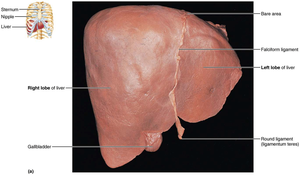
The liver is the largest gland in the body, weighing about 1.4 kg. It has four lobes (right, left, caudate, quadrate) and is separated by the falciform ligament. The liver produces about 900 ml of bile daily.
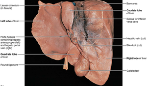
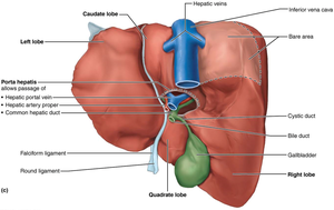
Bile Duct System: Left and right hepatic ducts join to form the common hepatic duct, which merges with the cystic duct from the gallbladder to form the bile duct, delivering bile to the duodenum.
Liver Histology
The liver is organized into hexagonal lobules with a central vein. Each lobule contains plates of hepatocytes, liver sinusoids, and a portal triad (branch of hepatic artery, portal vein, and bile duct). Blood flows toward the central vein, while bile flows in the opposite direction toward the bile duct.
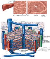
Stellate Macrophages: Located in sinusoids, these cells remove debris and old blood cells.
Composition and Function of Bile
Bile is a yellow-green, alkaline solution containing bile salts, bile pigments (mainly bilirubin), cholesterol, neutral fats, phospholipids, and electrolytes. Only bile salts and phospholipids aid in lipid digestion by emulsifying fats, making them more accessible to digestive enzymes. The main bile salts are cholic acid and chenodeoxycholic acid, which are amphipathic molecules (having both hydrophilic and hydrophobic regions).
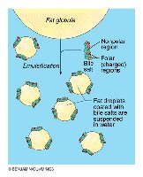
Bile Pigments: Bilirubin is converted to urobilinogen and stercobilin by intestinal bacteria, giving feces its color.
Enterohepatic Circulation: Bile salts are recycled from the intestine back to the liver, stimulating further bile production.
The Gallbladder
Structure and Function
The gallbladder is a thin-walled muscular sac that stores and concentrates bile by absorbing water and ions. Muscular contractions expel bile into the cystic duct, which joins the bile duct to deliver bile to the duodenum. Supersaturation of bile with cholesterol can lead to gallstone formation (biliary calculi).
The Pancreas
Structure and Function
The pancreas is mostly retroperitoneal, with its head encircled by the duodenum and its tail abutting the spleen. It produces 1200–1500 ml of pancreatic juice daily. The exocrine pancreas consists of acinar cells that produce digestive enzymes and duct cells that secrete bicarbonate to neutralize stomach acid.
Pancreatic Juice: Contains electrolytes (mainly bicarbonate), proteases (secreted in inactive form), amylase, lipases, and nucleases.
Activation of Proteases: Enteropeptidase, a brush border enzyme, activates trypsinogen to trypsin, which then activates other proteases.
Regulation of Bile and Pancreatic Secretion
Hormonal Control
During digestion, the hepatopancreatic sphincter relaxes in response to cholecystokinin (CCK), allowing bile and pancreatic juice to enter the duodenum. CCK is released in response to fatty chyme and stimulates gallbladder contraction, pancreatic enzyme secretion, and sphincter relaxation. Secretin stimulates the secretion of bicarbonate-rich fluid from the liver and pancreas.
The Small Intestine
Structure and Function
The small intestine is 6–7 meters long and extends from the pyloric sphincter to the ileocecal valve. It consists of three regions: duodenum, jejunum, and ileum. The small intestine is the primary site for digestion and absorption of nutrients.
Blood Supply: Supplied by the superior mesenteric artery and drained by the superior mesenteric vein into the hepatic portal vein.
Adaptations for Absorption
The small intestine has several modifications to increase surface area for absorption:
Circular folds: Permanent folds of mucosa and submucosa that slow the movement of chyme.
Villi: Finger-like projections that increase surface area, longest in the duodenum.
Microvilli: Cytoplasmic extensions forming the brush border, containing enzymes for final digestion steps.
Histology of the Small Intestine Wall
The mucosal epithelium contains five main cell types:
Enterocytes: Absorptive cells with microvilli; absorb nutrients and secrete intestinal juice.
Goblet cells: Produce mucus for lubrication and protection.
Enteroendocrine cells: Secrete hormones such as secretin and CCK.
Paneth cells: Secrete antimicrobial agents (defensins, lysozyme).
Stem cells: Continuously divide to replace epithelial cells.
Mucosa-associated lymphoid tissue (MALT), including Peyer's patches, provides immune protection. The submucosa contains areolar connective tissue and duodenal glands that secrete alkaline mucus. The muscularis externa consists of two layers of smooth muscle. About 1–2 liters of slightly alkaline intestinal juice are produced daily, mainly in response to hyperosmotic or acidic chyme.
