 Back
BackDigestive System: Pharynx, Esophagus, Stomach, and Accessory Organs
Study Guide - Smart Notes
 Tailored notes based on your materials, expanded with key definitions, examples, and context.
Pharynx and Esophagus
The Pharynx
The pharynx serves as a passageway for food, fluids, and air, connecting the mouth to the esophagus. It is lined with stratified squamous epithelium and contains mucus-producing glands for lubrication and protection. The external muscle layer consists of two skeletal muscle layers: an inner longitudinal layer and an outer pharyngeal constrictor layer, which encircle the wall of the pharynx and facilitate swallowing.
The Esophagus
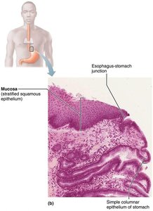
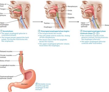
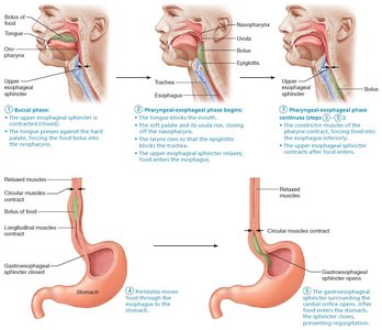
The esophagus is a flat, muscular tube that extends from the laryngopharynx to the stomach. It remains collapsed when not in use and passes through the diaphragm at the esophageal hiatus. The esophagus joins the stomach at the cardial orifice, which is surrounded by the gastroesophageal (cardiac) sphincter. This sphincter prevents acid reflux by keeping the orifice closed when food is not being swallowed. Mucus cells on both sides of the sphincter protect the esophagus from stomach acid.
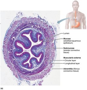
Microscopic Structure of the Esophagus
Mucosa: Stratified squamous epithelium (changes to simple columnar at the stomach)
Submucosa: Contains esophageal glands that secrete mucus to aid in bolus movement
Muscularis externa: Skeletal muscle superiorly, mixed in the middle, smooth muscle inferiorly
Adventitia: Outermost layer, composed of fibrous connective tissue (instead of serosa)
Deglutition (Swallowing)
Phases of Deglutition
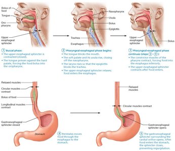
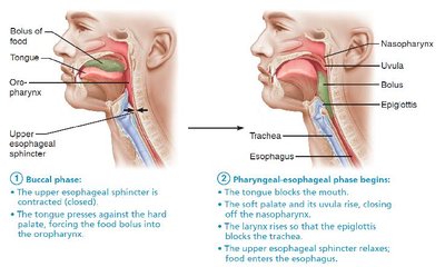
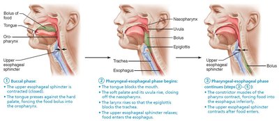
Deglutition, or swallowing, is the process of moving food from the mouth to the stomach. It involves the coordinated action of 22 muscle groups and occurs in two main phases:
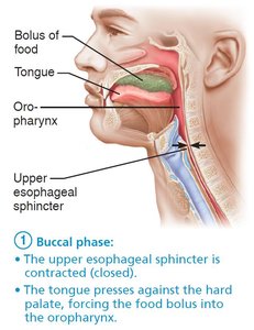
Buccal phase: Voluntary contraction of the tongue to push the bolus into the oropharynx.
Pharyngeal-esophageal phase: Involuntary phase controlled by the swallowing center in the medulla and lower pons, primarily involving the vagus nerve. This phase ensures the bolus passes through the pharynx and esophagus into the stomach.
The Stomach
Gross Anatomy of the Stomach
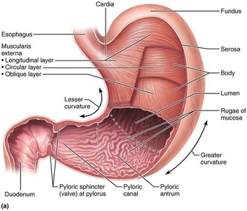
The stomach is a muscular, expandable organ that serves as a temporary storage tank and initiates the chemical breakdown of proteins. It converts the food bolus into a paste-like substance called chyme. The stomach can expand from about 50 mL when empty to up to 4 liters when full. The mucosa forms folds called rugae when the stomach is empty.
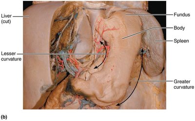
Major regions: Cardia, fundus, body, pyloric part (antrum, canal, pylorus)
Curvatures: Greater curvature (lateral), lesser curvature (medial)
Mesenteries: Lesser omentum (to liver), greater omentum (drapes over intestines)
Blood supply: Celiac trunk (gastric and splenic branches), hepatic portal system veins
Innervation: Sympathetic (thoracic splanchnic nerves via celiac plexus), parasympathetic (vagus nerve)
Microscopic Anatomy of the Stomach
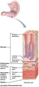
The stomach wall contains four tunics, with modifications in the muscularis and mucosa:
Muscularis externa: Three layers—longitudinal, circular, and oblique (unique to stomach)—allow for churning and mixing of chyme.
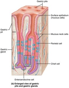
Mucosa: Simple columnar epithelium composed entirely of mucous cells, secreting a two-layer coat of alkaline mucus. The surface layer traps bicarbonate-rich fluid beneath it. The mucosa is dotted with gastric pits leading to gastric glands that produce gastric juice.
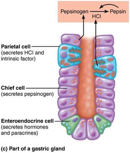
Types of Gland Cells
Mucous neck cells: Secrete thin, acidic mucus (function not fully understood).
Parietal cells: Secrete hydrochloric acid (HCl, pH 1.5–3.5) and intrinsic factor (for vitamin B12 absorption).
Chief cells: Secrete pepsinogen (inactive enzyme, activated to pepsin by HCl and pepsin) and gastric lipases.
Enteroendocrine cells: Secrete chemical messengers (serotonin, histamine, somatostatin, gastrin) into the lamina propria.
Mucosal Barrier
The stomach is protected from harsh digestive conditions by a mucosal barrier consisting of:
Thick layer of bicarbonate-rich mucus
Tight junctions between epithelial cells
Rapid replacement of damaged epithelial cells (every 3–6 days)
Clinical Correlations
Heartburn and GERD
Heartburn is caused by stomach acid regurgitating into the esophagus, often due to a weak gastroesophageal sphincter. It is the first symptom of gastroesophageal reflux disease (GERD) and can be triggered by overeating, obesity, pregnancy, or hiatal hernia. Chronic exposure can lead to esophagitis, ulcers, or cancer.
Gastritis and Peptic Ulcers
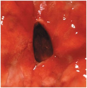
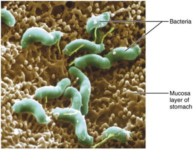
Gastritis is inflammation of the stomach mucosa, often caused by breach of the mucosal barrier. Peptic ulcers are erosions in the stomach wall, most commonly caused by Helicobacter pylori infection or NSAIDs (e.g., aspirin). Severe ulcers can lead to peritonitis or hemorrhage.
Digestive Processes in the Stomach
Breakdown of food and holding area for chyme
Delivers chyme to small intestine
Denatures proteins with HCl
Pepsin digests proteins; in infants, rennin digests milk protein (casein)
Absorbs some lipid-soluble substances (alcohol, aspirin)
Secretion of intrinsic factor (essential for vitamin B12 absorption and red blood cell maturation)
Regulation of Gastric Secretion
Phases of Gastric Secretion
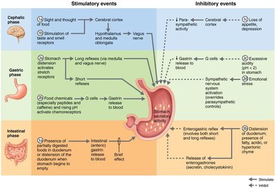
Gastric secretion is regulated by neural (vagus nerve, sympathetic inhibition) and hormonal (gastrin) mechanisms. There are three phases:
Cephalic (reflex) phase: Triggered by sight, smell, taste, or thought of food.
Gastric phase: Lasts 3–4 hours, provides most gastric juice. Stimulated by stomach distension and food chemicals (proteins, caffeine, low acidity) activating G cells to secrete gastrin.
Intestinal phase: Brief stimulation as chyme enters duodenum, followed by inhibition to prevent overfilling and excessive acidity.
Hormones and Paracrines in Digestion
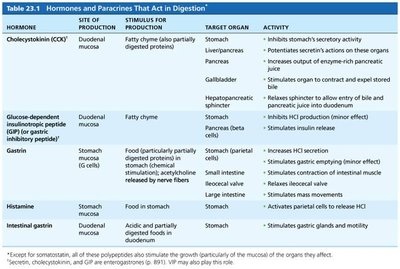
Hormone | Site of Production | Stimulus | Target Organ | Activity |
|---|---|---|---|---|
Cholecystokinin (CCK) | Duodenal mucosa | Fatty chyme | Stomach, liver/pancreas, gallbladder | Inhibits gastric secretion, stimulates enzyme-rich pancreatic juice, stimulates gallbladder contraction |
Gastrin | Stomach (G cells) | Food in stomach | Stomach, small intestine, ileocecal valve, large intestine | Increases HCl secretion, stimulates gastric emptying, stimulates contraction of intestinal muscle |
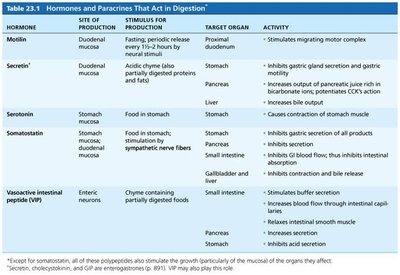
Secretin | Duodenal mucosa | Acidic chyme | Stomach, pancreas, liver | Inhibits gastric secretion, increases bile output, increases pancreatic bicarbonate output |
Histamine | Stomach mucosa | Food in stomach | Stomach | Activates parietal cells to release HCl |
Somatostatin | Stomach, duodenal mucosa, pancreas | Food in stomach, sympathetic nerve fibers | Stomach, pancreas, small intestine, liver | Inhibits gastric secretion, inhibits secretion of pancreatic hormones, inhibits GI blood flow |
Mechanism of HCl Formation
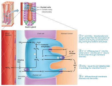
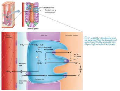
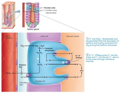
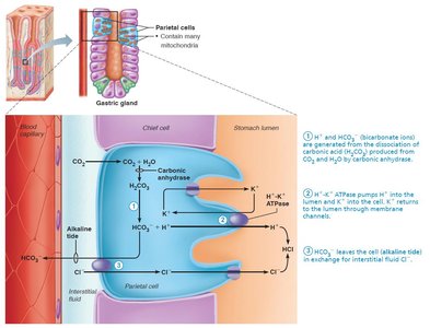
Parietal cells secrete HCl into the stomach lumen via the following mechanism:
H+ ions are generated from the dissociation of carbonic acid (H2CO3), produced from CO2 and H2O by carbonic anhydrase.
H+ is pumped into the lumen via H+/K+ ATPase (proton pump).
HCO3− is exported to the blood in exchange for Cl− (alkaline tide).
Cl− diffuses into the lumen and combines with H+ to form HCl.
Summary Table: Layers of the Alimentary Canal
Layer | Location | Function |
|---|---|---|
Mucosa | Innermost | Secretion, absorption, protection |
Submucosa | External to mucosa | Blood vessels, nerves, lymphatics |
Muscularis externa | External to submucosa | Segmentation and peristalsis |
Serosa/Adventitia | Outermost | Protection, structural support |
Additional info: The above notes integrate and expand upon the provided content, ensuring a comprehensive, exam-ready summary for Anatomy & Physiology students.
