 Back
BackDigestive System: Structure and Function (Anatomy & Physiology Study Guide)
Study Guide - Smart Notes
 Tailored notes based on your materials, expanded with key definitions, examples, and context.
Digestive System Overview
Introduction to the Digestive System
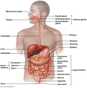
The digestive system is responsible for the breakdown and absorption of nutrients from food, as well as the elimination of waste products. It consists of the digestive tract (also known as the gastrointestinal tract or alimentary canal) and accessory organs that assist in digestion.
Digestive Tract: Extends from the mouth to the anus (~9 meters).
Accessory Organs: Include salivary glands, teeth, tongue, liver, gallbladder, and pancreas.
Major Functions of the Digestive System
The digestive system performs several essential functions to maintain homeostasis and provide energy for the body.
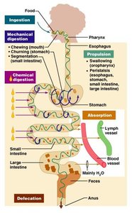
Ingestion: Intake of food through the mouth.
Mechanical Digestion: Physical breakdown of food by chewing, churning, and segmentation.
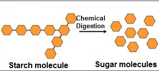
Chemical Digestion: Enzymatic breakdown of food into smaller molecules.
Secretion: Release of digestive substances into the lumen.
Absorption: Movement of nutrients into blood or lymph.
Excretion: Removal of waste products via defecation.
Histology of the Digestive Tract
Layers of the Digestive Tract
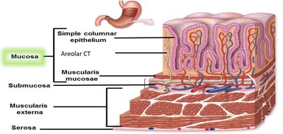
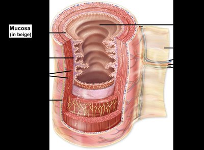
The wall of the digestive tract is composed of four main layers, each with distinct functions and structures.
Mucosa: Innermost layer; includes epithelium, lamina propria, and muscularis mucosae.
Submucosa: Connective tissue containing blood vessels, lymphatics, nerves, and glands.
Muscularis Externa: Smooth muscle responsible for peristalsis and segmentation; typically two layers (circular and longitudinal), with a third layer in the stomach.
Adventitia/Serosa: Outermost layer; adventitia for retroperitoneal organs, serosa for intraperitoneal organs.
Mucosa and Submucosa Details
The mucosa varies by location, with nonkeratinized stratified squamous epithelium in the mouth, pharynx, esophagus, and anus, and simple columnar epithelium in the stomach and intestines. The submucosa supports the mucosa and contains essential vessels and nerves.
Muscularis Externa and Peristalsis
The muscularis externa is responsible for the movement of food through the digestive tract via peristalsis, which are rhythmic contractions of smooth muscle.

Serosa, Adventitia, and Mesenteries
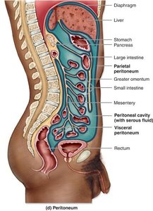
Serosa is a serous membrane covering intraperitoneal organs, while adventitia is a connective tissue layer for retroperitoneal organs. Mesenteries are double layers of peritoneum that attach organs to the cavity wall, store fat, and hold blood vessels and nerves.
Components of the Digestive Tract
Oral Cavity
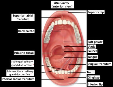
The oral cavity is the entry point for food and is involved in mechanical digestion, lubrication, and the initial chemical digestion of carbohydrates. It forms the bolus for swallowing.
Pharynx
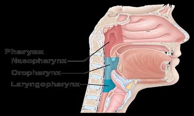
The pharynx connects the oral cavity to the esophagus and is divided into three regions: nasopharynx, oropharynx, and laryngopharynx. The oropharynx and laryngopharynx are part of the digestive system and move the bolus to the esophagus.
Esophagus
The esophagus is a muscular tube posterior to the trachea, extending from the laryngopharynx to the stomach. It uses peristalsis to move solids and liquids from the pharynx to the stomach. The cardiac/gastroesophageal sphincter regulates entry into the stomach.
Stomach
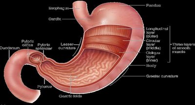
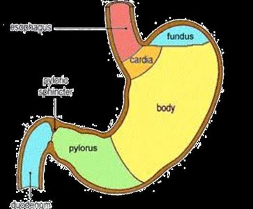
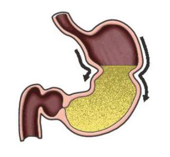
The stomach is a J-shaped organ that stores ingested food, performs mechanical digestion (churning), and initiates chemical digestion of proteins. It is divided into four regions: cardia, fundus, body, and pyloric region. The pyloric sphincter controls the movement of chyme into the duodenum.
Mucosa: Contains gastric pits and glands that secrete mucus, hormones, pepsinogen, HCl, intrinsic factor, and buffers.
Muscularis mucosa: Forms gastric folds (rugae) to increase surface area.
Small Intestine
The small intestine is the primary site for digestion and absorption. It consists of three segments: duodenum (closest to stomach), jejunum, and ileum. The mucosa has villi and microvilli to increase surface area for absorption.
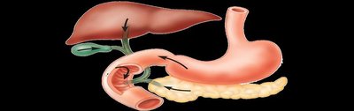
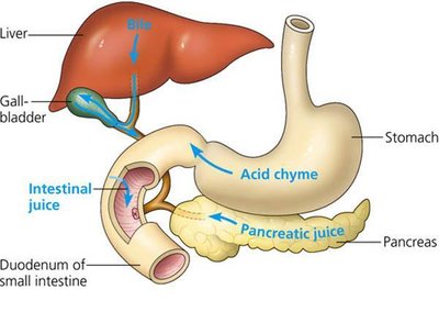
Duodenum: Receives chyme from the stomach and digestive juices from the liver, gallbladder, and pancreas.
Jejunum: Main site for nutrient absorption.
Ileum: Absorbs remaining nutrients and connects to the large intestine.
Large Intestine
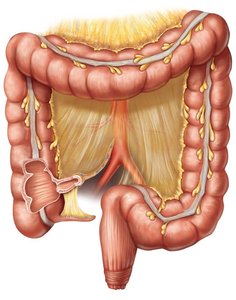
The large intestine absorbs water, vitamins, and compacts feces. It consists of the cecum, colon (ascending, transverse, descending, sigmoid), rectum, and anal canal. The rectum is the last 20 cm of the colon and stores solid waste before defecation.
Cecum: Blind pouch below the ileum; appendix attached.
Colon: Main site for water and vitamin absorption.
Rectum: Stores fecal material; lacks serosa.
Anal canal: Forms internal (involuntary) and external (voluntary) anal sphincters.
Accessory Organs
Accessory organs assist in digestion by producing and releasing substances necessary for the breakdown and absorption of nutrients.
Salivary glands: Produce saliva for lubrication and initial carbohydrate digestion.
Liver: Produces bile for lipid digestion.
Gallbladder: Stores and releases bile.
Pancreas: Produces digestive enzymes and bicarbonate.
Summary Table: Layers of the Digestive Tract
Layer | Main Components | Function |
|---|---|---|
Mucosa | Epithelium, lamina propria, muscularis mucosae | Secretion, absorption, protection |
Submucosa | Connective tissue, blood vessels, nerves, glands | Support, transport, innervation |
Muscularis Externa | Smooth muscle (circular, longitudinal, oblique in stomach) | Peristalsis, segmentation |
Serosa/Adventitia | Serous membrane or connective tissue | Protection, anchoring |
Key Terms and Definitions
Peristalsis: Rhythmic contractions of smooth muscle that propel food through the digestive tract.
Chyme: Semi-fluid mass of partially digested food expelled by the stomach into the duodenum.
Bile: Fluid produced by the liver and stored in the gallbladder, essential for emulsifying fats.
Bolus: Chewed food mixed with saliva, ready for swallowing.
Haustra: Pouches in the colon formed by the contraction of teniae coli.
Example: Chemical Digestion of Starch
Starch molecules are broken down into sugar molecules by enzymes during chemical digestion, facilitating absorption in the small intestine.
Additional info:
Most nutrients absorbed in the small intestine enter the hepatic portal system and are processed by the liver before entering systemic circulation.
The appendix contains lymphoid tissue and may play a role in immune function.
