 Back
BackDigestive System Structure and Function: ANP Study Notes
Study Guide - Smart Notes
 Tailored notes based on your materials, expanded with key definitions, examples, and context.
Digestive System Overview
Major Organs and Accessory Organs
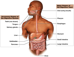
The digestive system is a long, muscular tube (the digestive tract) with specialized regions and accessory organs that work together to process food, absorb nutrients, and eliminate waste. The tract is lined with ridges and folds to increase surface area for absorption, supporting cell maintenance and growth.
Major organs: Oral cavity, pharynx, esophagus, stomach, small intestine, large intestine, anus
Accessory organs: Teeth, tongue, salivary glands, liver, gallbladder, pancreas
The enteric nervous system is a network of sensory neurons and interneurons within the digestive system, controlling most digestive activities. It is influenced by the autonomic nervous system (ANS):
Parasympathetic stimulation: Increases digestive activity
Sympathetic stimulation: Decreases digestive activity
Structure of the Digestive Tract
Four Generic Layers (MSMS)
The digestive tract wall is organized into four main layers, each with distinct functions:
Mucosa (inner lining): Mucous membrane moistened by glandular secretions; contains the lamina propria (areolar tissue). The specific cell type varies by location.
Submucosa: Dense connective tissue with blood vessels, lymphatics, nerve plexus, and exocrine glands. In some regions, it secretes buffers and enzymes.
Muscular layer: Smooth muscle in two layers (inner circular, outer longitudinal) for mechanical processing and movement. The stomach has an additional oblique layer.
Serosa: Visceral peritoneum covering most of the tract in the abdominal cavity. In the oral cavity, pharynx, esophagus, and rectum, this is replaced by the adventitia (collagen fibers).
Mucosa histology:
Stratified squamous epithelium at the beginning and end of the tract (for abrasion resistance)
Simple columnar epithelium with goblet cells in the stomach, small, and large intestines (for secretion and absorption)
Villi: Small mucosal projections in the small intestine that increase surface area for absorption
Smooth Muscle and Motility
Smooth Muscle in the Digestive Tract
Smooth muscle cells in the digestive tract walls are electrically connected by gap junctions and mechanically by dense bodies, allowing them to contract as a single unit. Stimulation can be neural, hormonal, or chemical, and some regions are rhythmically stimulated by pacesetter cells.
Peristalsis: Propulsion of Food
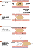
Peristalsis is a wave-like muscle contraction that propels food (bolus) along the digestive tract. The process involves:
Circular muscles contracting behind the bolus
Longitudinal muscles ahead of the bolus contracting
Sequential contraction of circular muscle layer forces the bolus forward
Segmentation: Mixing of Contents
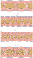
Segmentation involves cycles of contraction by the inner circular muscle, churning and mixing the contents in the small intestine without forward movement. This process fragments the bolus and mixes it with intestinal secretions, enhancing digestion and absorption.
No set pattern of contractions; no particular direction of movement
Occurs mainly in the small intestine and some parts of the large intestine
Regulation of Digestive Activities
Chemical and Physical Factors
Digestive activities are regulated by various factors at specific tract locations:
pH changes in the lumen
Physical distortion of the digestive tract wall
Nutrients or chemical messengers released by the mucosa
Neural (Reflex) Control
Short reflexes (myenteric reflexes): Triggered by chemoreceptors or stretch receptors in the tract wall; controlled by neurons in the myenteric plexus
Long reflexes: Involve interneurons and motor neurons of the CNS; control large-scale peristalsis and may involve parasympathetic fibers synapsing in the myenteric plexus
Hormonal Control Mechanisms
Multiple hormones produced by enteroendocrine cells in the digestive tract epithelium regulate digestive function, coordinating secretion and motility.
Oral Cavity, Teeth, and Swallowing
Oral Cavity Structure and Function
The oral cavity is lined by stratified squamous epithelium for abrasion resistance. Keratinized areas (superior tongue, hard palate) resist severe abrasion, while nonkeratinized areas (cheeks, lips, inferior tongue) are more delicate. Thin mucosa under the tongue allows rapid absorption of lipid-soluble drugs (e.g., nitroglycerin). Carbohydrate and lipid digestion begins here, but no nutrient absorption occurs.
Teeth and Supporting Structures
Gingival sulcus: Epithelial attachment blocks bacteria from deeper tissues
Periodontal ligament: Connects root dentin to alveolar bone (gomphosis articulation)
Root canal: Passageway for blood vessels and nerves to the pulp cavity
Clinical note: Infections in the root canal can lead to sepsis.
Pharynx and Esophagus: Swallowing (Deglutition)
Swallowing occurs in three phases:
Buccal phase (voluntary): Tongue pushes bolus into oropharynx; soft palate elevates to seal nasopharynx
Pharyngeal phase: Pharyngeal muscles contract, larynx elevates, epiglottis folds, and bolus moves into esophagus
Esophageal phase: Bolus triggers opening of lower esophageal sphincter (LES) and enters the stomach
Mesenteries and Stomach Structure
Mesenteries
Greater omentum: Fatty tissue attached to stomach and transverse colon; provides padding and protection
Lesser omentum: Connects stomach to liver; provides access for blood vessels and other structures
Stomach Regions and Wall Structure
Fundus: Superior region
Cardia: Borders gastroesophageal junction; secretes mucus to protect esophagus
Body: Main mixing area
Pylorus: Sharp curve; changes shape with mixing movements
The stomach wall has three layers of smooth muscle (circular, longitudinal, oblique) to strengthen the wall and assist in mixing and churning food into chyme. Rugae (temporary mucosal folds) allow the stomach to expand.
Cardiac sphincter (LES): Controls entry from esophagus
Pyloric sphincter: Regulates release of chyme into the duodenum
Stomach Functions and Gastric Glands
Functions of the Stomach
Temporary storage of ingested food
Mechanical digestion (mixing and churning)
Chemical digestion (acid and enzymes)
Production of intrinsic factor (for vitamin B12 absorption)
Stomach Wall and Gastric Glands
The mucosa (inner layer) is simple columnar epithelium, producing alkaline mucus to protect against acid and enzymes. Gastric epithelial cells have a short lifespan (3–7 days).
Gastric glands: Secrete most of the acid (HCl) and enzymes for digestion; dominated by parietal and chief cells
Pyloric glands: Secrete mucus and hormones to coordinate digestive activity
Cells of Gastric Glands
Parietal cells: Secrete intrinsic factor (for B12 absorption) and hydrochloric acid (HCl) to maintain low stomach pH (1.5–2.0) for protein digestion
Chief cells: Secrete pepsinogen, which is activated by HCl to form pepsin (digests proteins)
G cells (enteroendocrine cells): Secrete hormones such as gastrin to regulate stomach acid
HCl Production
Hydrochloric acid is produced by the following process:
H+ ions from carbonic acid (H2CO3) enter the lumen
Cl- ions from interstitial fluid also enter the lumen
H+ and Cl- combine in the lumen to form HCl
(from parietal cell) (from interstitial fluid) (in gastric lumen)
