 Back
BackDigestive System: Structure and Function (ANP1115 Lecture 10 Study Notes)
Study Guide - Smart Notes
 Tailored notes based on your materials, expanded with key definitions, examples, and context.
Digestive System Overview
General Anatomical Features
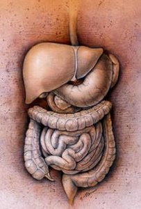
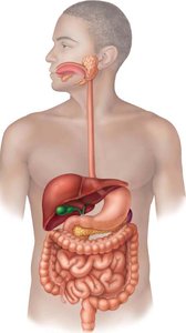
The digestive system is responsible for providing the body with a steady supply of nutrients, water, and inorganic ions. It consists of the alimentary canal (gastrointestinal tract) and accessory digestive organs. The tract itself is a continuous tube open at both ends, making its lumen continuous with the external environment.
Main organs: Mouth, pharynx, esophagus, stomach, small intestine, large intestine
Accessory structures: Teeth, tongue, salivary glands, pancreas, liver, gallbladder
Accessory organs assist with mechanical digestion or secrete substances (e.g., saliva, bile, enzymes) to aid digestion.
Major Digestive Processes
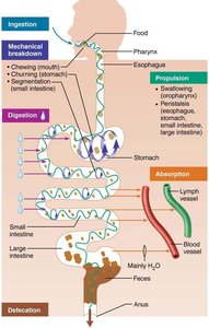
Six key processes occur in the digestive system, each with specific locations and functions:
Ingestion: Taking food into the mouth
Propulsion: Moving food through the tract (includes swallowing and peristalsis)
Mechanical breakdown: Chewing, churning, and segmentation to increase surface area
Digestion: Enzymatic breakdown of food into absorbable molecules
Absorption: Transport of digested end products into blood or lymph
Defecation: Elimination of indigestible substances as feces
Motility Patterns
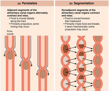
Two primary motility patterns move and mix food in the digestive tract:
Peristalsis: Adjacent segments of the tract alternately contract and relax, propelling food distally.
Segmentation: Nonadjacent segments contract and relax, mixing food and breaking it down mechanically.
Peritoneum and Mesenteries
Peritoneum
The peritoneum is a double-layered serous membrane associated with the abdominal organs:
Visceral peritoneum: Covers external surfaces of digestive organs
Parietal peritoneum: Lines the body wall of the abdominopelvic cavity
The space between layers contains serous fluid to reduce friction
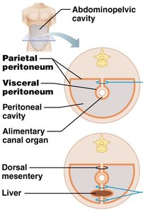
Mesenteries
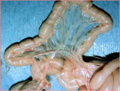
A mesentery is a double layer of peritoneum that extends from the body wall to digestive organs. It:
Provides a route for blood vessels, lymphatics, and nerves
Holds organs in place
Stores fat
Retroperitoneal and Intraperitoneal Organs
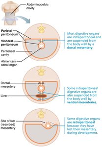
Most digestive organs are intraperitoneal (suspended by mesenteries), but some (e.g., pancreas, duodenum) are retroperitoneal, having lost their mesentery during development.
Blood Supply and Splanchnic Circulation
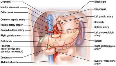
The digestive system requires an extensive blood supply for nutrient absorption and transport. The splanchnic circulation includes arteries branching from the aorta (hepatic, splenic, left gastric, mesenteric arteries) and the hepatic portal circulation, which delivers nutrient-rich blood from the digestive organs to the liver for processing.
Histological Organization of the Alimentary Canal
Four Basic Layers (Tunics)
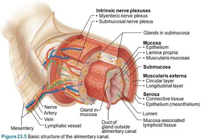
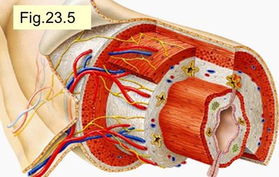
The wall of the alimentary canal consists of four main layers:
Mucosa: Innermost layer; functions in secretion, absorption, and protection
Submucosa: Connective tissue with blood vessels, lymphatics, nerves, and glands
Muscularis externa: Smooth muscle responsible for peristalsis and segmentation
Serosa: Outermost layer; visceral peritoneum (or adventitia in some regions)
Mucosa
Epithelium: Stratified squamous (mouth, esophagus, anus) or simple columnar (stomach, intestines); contains goblet cells that secrete mucus
Lamina propria: Loose connective tissue with capillaries and MALT (mucosa-associated lymphoid tissue)
Muscularis mucosae: Thin layer of smooth muscle for local movements
Submucosa
Dense connective tissue with blood and lymphatic vessels, nerves, and glands
Contains the submucosal plexus (nerve network regulating glands and smooth muscle)
Muscularis Externa
Typically two layers: inner circular and outer longitudinal smooth muscle (stomach has an additional oblique layer)
Responsible for peristalsis and segmentation
Contains the myenteric plexus (nerve network controlling motility)
Thickened regions form sphincters to control passage of materials
Serosa
Outer connective tissue covering (visceral peritoneum)
In the esophagus and retroperitoneal organs, this layer is called the adventitia
Neural Control of Digestive Function
Enteric Nervous System
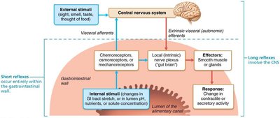
The digestive tract has its own nervous system, the enteric nervous system, consisting of the submucosal and myenteric plexuses. It regulates secretory activity, motility, and blood flow through both short (local) and long (CNS-mediated) reflexes.
The Mouth and Associated Structures
Anatomy of the Oral Cavity
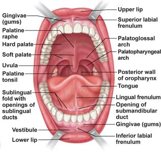
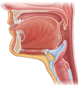
The mouth (oral cavity) is the entry point for food and is lined with stratified squamous epithelium for protection. The roof is formed by the hard and soft palates, and the floor by the tongue. The soft palate rises during swallowing to close off the nasopharynx.
Lips and cheeks: Contain skeletal muscle (orbicularis oris, buccinators) to hold food and aid speech
Labial and lingual frenula: Folds of tissue connecting lips and tongue to the gums
The Tongue
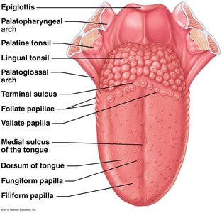
The tongue is a muscular organ essential for manipulating food, mixing it with saliva, and forming a bolus for swallowing. It contains intrinsic muscles (change shape) and extrinsic muscles (change position).
Papillae and Taste Buds
Filiform papillae: Conical, keratinized, provide friction
Fungiform papillae: Mushroom-shaped, contain taste buds
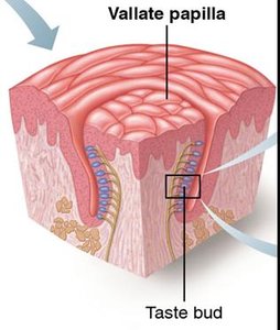
Vallate papillae: Large, V-shaped row at back of tongue, contain taste buds
Foliate papillae: On sides of tongue, contain taste buds
Salivary Glands and Saliva
Types and Functions of Salivary Glands
There are three pairs of major (extrinsic) salivary glands and numerous minor (intrinsic) glands:
Parotid glands: Mostly serous cells (watery secretion)
Submandibular glands: Mixed serous and mucus cells
Sublingual glands: Primarily mucus cells
Intrinsic buccal glands keep the mouth moist between meals
Composition and Function of Saliva
Mostly water (97–99.5%), slightly acidic (pH 6.75–7.00)
Contains electrolytes, salivary amylase, lingual lipase, mucin, lysozyme, IgA, and metabolic wastes
Functions: cleanses mouth, dissolves food for taste, moistens and compacts food, begins starch digestion
Control of Salivation
Stimulated by parasympathetic nervous system (cranial nerves VII and IX)
Inhibited by sympathetic stimulation and dehydration (causing dry mouth)
Teeth and Mastication
Dentition and Dental Formula
Humans have two sets of teeth:
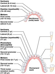
Primary (deciduous) teeth: 20, erupt between 6–24 months
Permanent teeth: 32, replace primary teeth between ages 6–12
Teeth are classified as incisors, canines, premolars, and molars based on shape and function
Dentition | Dental Formula (one side) | Total Teeth |
|---|---|---|
Primary | 2I, 1C, 2M | 20 |
Permanent | 2I, 1C, 2PM, 3M | 32 |
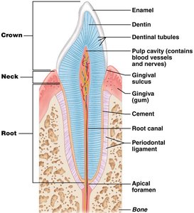
Tooth Structure
Crown: Exposed part, covered by enamel (hardest substance in body)
Root: Embedded in jawbone, covered by cement, attached to periodontal ligament
Dentin: Bone-like material under enamel, maintained by odontoblasts
Pulp cavity: Contains blood vessels and nerves
Deglutition (Swallowing) and the Esophagus
Deglutition
Swallowing involves coordinated activity of 22 muscle groups and two main phases:
Buccal phase: Voluntary, tongue pushes bolus into pharynx
Pharyngeal-esophageal phase: Involuntary, controlled by the swallowing center in the medulla and lower pons, moves food through pharynx and esophagus to stomach
The Esophagus
Muscular tube (~25 cm), collapsed when empty
Transitions from skeletal to smooth muscle along its length
Joins stomach at the gastroesophageal sphincter
Lined with stratified squamous epithelium for protection
Stomach: Structure and Function
Functional Anatomy
The stomach acts as a temporary storage tank, initiates protein digestion, and converts food to chyme. It is divided into regions: cardia, fundus, body, and pyloric region. The stomach wall contains rugae (folds) that allow expansion.
Microscopic Anatomy
Surface epithelium: Goblet cells produce mucus and a bicarbonate-rich layer for protection
Gastric pits: Lead to gastric glands that secrete gastric juice
Cell types:
Mucous neck cells: Secrete acidic mucin
Parietal cells: Secrete HCl (activates pepsinogen, denatures proteins) and intrinsic factor (vitamin B12 absorption)
Chief cells: Secrete pepsinogen (precursor to pepsin) and gastric lipase
Enteroendocrine cells: Secrete hormones (gastrin, histamine, serotonin, somatostatin)
Protection of the Stomach
Thick, bicarbonate-rich mucus layer
Tight junctions between epithelial cells
Rapid epithelial cell turnover (renewed every 3–6 days by stem cells)
