 Back
BackDigestive System: Structure and Function of the Alimentary Canal
Study Guide - Smart Notes
 Tailored notes based on your materials, expanded with key definitions, examples, and context.
Oral Cavity
Anatomy and Boundaries
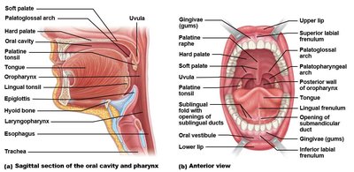
The oral cavity, or mouth, is the entryway to the digestive tract and is bounded by the lips, cheeks, palate, and tongue. The space between the lips/cheeks and teeth is called the vestibule. The lips surround the oral orifice and are connected to the gums by the labial frenulum. The roof of the oral cavity consists of the hard palate (formed by the maxillae and palatine bones) and the soft palate (muscular, closes off the nasopharynx during swallowing).
Tongue
Structure and Function
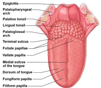
The tongue is composed of skeletal muscle and connective tissue, allowing for a wide range of movements. Intrinsic muscles change the shape of the tongue, while extrinsic muscles alter its position. The surface of the tongue contains papillae (not taste buds themselves), with taste buds located in grooves between papillae. There are four types of papillae: vallate, fungiform, filiform, and foliate.
Teeth
Dental Formulae and Types
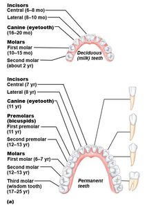
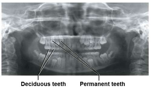
Humans have two sets of teeth: deciduous (milk) teeth and permanent teeth. Deciduous teeth erupt from about 6 to 24 months of age (20 teeth: 2 incisors, 1 canine, 2 molars per quadrant). Permanent teeth erupt from 6-12 years, with third molars (wisdom teeth) appearing between 17-25 years (32 teeth: 2 incisors, 1 canine, 2 premolars, 3 molars per quadrant).
Tooth Structure and Classification
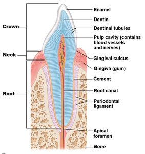
Teeth are classified by shape: incisors, canines, premolars, and molars. The occlusion refers to the fit of upper and lower teeth. Tooth structure (superficial to deep): enamel (inorganic, hardest substance in the body), dentin (organic), and pulp cavity (contains nerves and blood vessels). Each tooth has three regions: crown, neck, and root.
Salivary Glands
Types and Locations
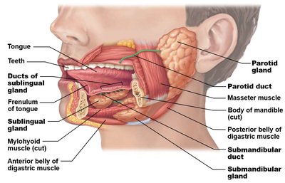
Salivary glands are classified as intrinsic (minor, scattered throughout the oral cavity) and extrinsic (major, paired glands). The three major extrinsic glands are:
Parotid gland: Largest, located near the ear, opens lateral to the second upper molar.
Sublingual gland: Inferior to the tongue, opens directly superior to the gland.
Submandibular gland: Medial to the mandible, opens lateral to the lingual frenulum.
Esophagus
Structure and Sphincters
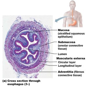
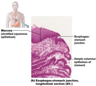
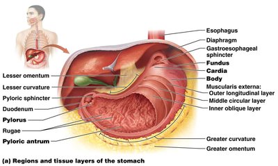
The esophagus is a muscular tube connecting the pharynx to the stomach. Its muscularis layer varies: upper third is skeletal muscle, middle third is mixed, and lower third is smooth muscle. The pharyngoesophageal sphincter regulates entry from the pharynx, and the gastroesophageal (cardiac) sphincter controls entry into the stomach. The esophagus passes through the diaphragm at the esophageal hiatus and enters the stomach at the cardinal orifice.
Stomach
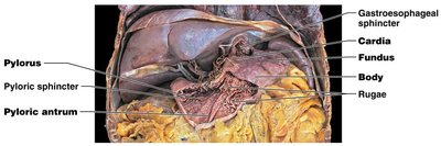
Regions and Curvatures
The stomach is a muscular organ with four main regions: cardiac (junction with esophagus), fundus (superior dome), body (largest region), and pyloric (junction with duodenum). The greater curvature is anterior, inferior, and left lateral, while the lesser curvature is superior and medial. The stomach is attached to the greater omentum and lesser omentum.
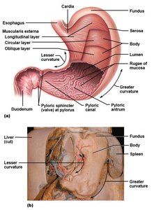
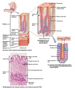
Histology and Function
The mucosal layer of the stomach is modified into gastric pits (ducts) and gastric glands (secretory). The length and shape of pits and glands vary by region. The muscularis layer has three smooth muscle layers: circular, longitudinal, and oblique, aiding in mechanical digestion. Rugae are gastric folds that allow for stomach expansion. The pyloric sphincter regulates the passage of chyme into the duodenum.
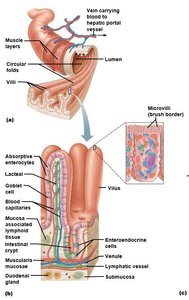
Small Intestine
Structure and Specializations
The small intestine extends from the pyloric sphincter to the ileocecal valve. Its mucosal and submucosal layers are highly modified for absorption:
Plicae circulares: Permanent folds of mucosa and submucosa.
Villi: Fingerlike projections of the mucosa, increasing surface area.
Microvilli: Folds of the apical surface of enterocytes, forming the brush border.
Intestinal glands (crypts): Invaginations into the lamina propria.
Duodenal (Brunner’s) glands: Present in the submucosa of the duodenum, secrete alkaline mucus.
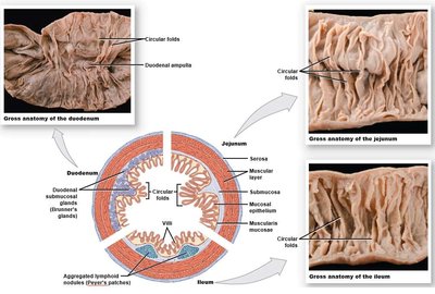
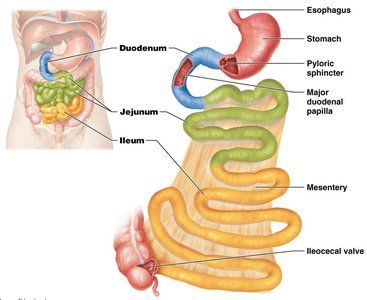
Duodenum, Jejunum, and Ileum
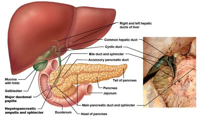
The duodenum is the first segment (C-shaped, retroperitoneal), receiving digestive enzymes and bile via the hepatopancreatic ampulla at the major duodenal papilla. The jejunum (40% of length) and ileum (55% of length) are intraperitoneal; the ileum contains Peyer’s patches (lymph nodules) in the mucosa.
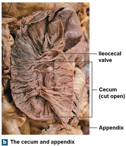
Ileocecal Valve and Appendix
Structure and Function
The ileocecal valve is a raised mucosal fold between the ileum and cecum, regulating passage of intestinal contents. The appendix is a blind-ended tube with abundant lymph nodules in its submucosa, located posterior-medial to the cecum.
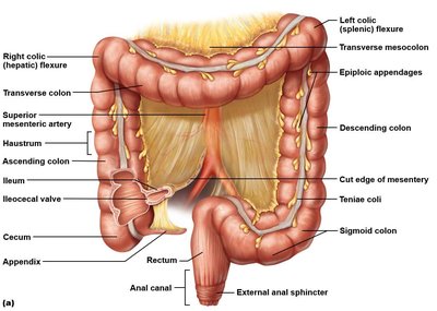
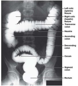
Large Intestine
Regions and Special Features
The large intestine is approximately 1.5 meters long and consists of the cecum, colon (ascending, transverse, descending, sigmoid), rectum, and anus. Special features include:
Teniae coli: Three bands of longitudinal muscle creating haustra (pouches).
Epiploic appendages: Fat-filled pouches of visceral peritoneum.
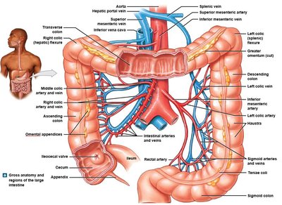
Anatomical Landmarks and Segments
The ascending colon runs along the right posterior wall, the transverse colon is intraperitoneal, the descending colon is on the left posterior wall, and the sigmoid colon enters the true pelvis. The rectum is secondarily retroperitoneal, and the anus is external to the abdominopelvic cavity.

Tunics of the Large Intestine
Histological Features
The mucosal layer of the large intestine lacks villi but contains abundant goblet cells for mucus production and numerous lymph nodules. The muscular layer consists of three layers: circular, longitudinal, and oblique (in some regions).
Rectum and Anus
Structure and Sphincters
The rectum contains three transverse folds and lacks teniae coli. The anal canal is maintained by the levator ani muscle and contains anal columns and valves. Anal sinuses secrete mucus during defecation. The internal anal sphincter is smooth muscle (involuntary), while the external anal sphincter is skeletal muscle (voluntary). Both are contracted at rest; relaxation of the internal sphincter is triggered by rectal stretch, and voluntary control of the external sphincter allows for defecation.
Region | Key Features |
|---|---|
Oral Cavity | Lips, palate, tongue, teeth, salivary glands |
Esophagus | Muscular tube, sphincters, tissue layers |
Stomach | Four regions, rugae, gastric glands, three muscle layers |
Small Intestine | Plicae circulares, villi, microvilli, duodenal glands, Peyer’s patches |
Large Intestine | Teniae coli, haustra, epiploic appendages, goblet cells |
Rectum & Anus | Transverse folds, anal columns, internal/external sphincters |
