 Back
BackDigestive System: Structure, Function, and Associated Organs
Study Guide - Smart Notes
 Tailored notes based on your materials, expanded with key definitions, examples, and context.
Digestive System Overview
Main Functions of the Digestive System
The digestive system is responsible for processing food, extracting nutrients, and eliminating waste. Its primary functions include:
Ingestion: Taking in food through the mouth.
Mechanical and Chemical Breakdown: Chewing, mixing, and enzymatic digestion of food.
Absorption: Uptake of nutrient molecules into the bloodstream or lymphatic system.
Defecation: Removal of indigestible remains from the body.
Organs of the Digestive System
The digestive system consists of two main groups of organs:
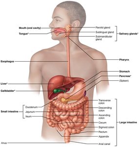
Alimentary Canal (GI Tract): A continuous muscular tube from mouth to anus, including the mouth, pharynx, esophagus, stomach, small intestine, large intestine, and anus. It digests and absorbs food.
Accessory Digestive Organs: These include the teeth, tongue, gallbladder, and digestive glands (salivary glands, liver, pancreas) that produce secretions aiding digestion.
Digestive Processes
Essential Activities of Digestion
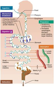
Food processing in the digestive system involves six key activities:
Ingestion: Eating food.
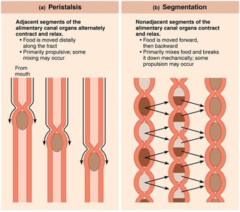
Propulsion: Moving food through the alimentary canal via swallowing and peristalsis (alternating waves of contraction and relaxation).
Mechanical Breakdown: Chewing, mixing with saliva, churning in the stomach, and segmentation (local constriction in the intestine).
Digestion: Enzymatic breakdown of complex molecules into simpler building blocks.
Absorption: Passage of digested fragments into blood or lymph.
Defecation: Elimination of indigestible substances as feces.
Organization and Anatomy of the Digestive System
Peritoneum and Mesenteries
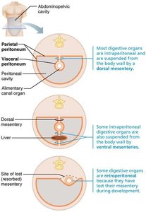
The peritoneum is a serous membrane lining the abdominal cavity, consisting of:
Visceral Peritoneum: Covers external surfaces of most digestive organs.
Parietal Peritoneum: Lines the body wall.
Peritoneal Cavity: Fluid-filled space between the two peritoneums, lubricating organs.
Mesentery is a double layer of peritoneum that provides routes for blood vessels, lymphatics, and nerves, holds organs in place, and stores fat.
Intraperitoneal Organs: Located within the peritoneum.
Retroperitoneal Organs: Located outside or posterior to the peritoneum (e.g., most of the pancreas, duodenum, parts of the large intestine).
Clinical Note: Peritonitis
Peritonitis: Inflammation of the peritoneum, often caused by abdominal wounds, perforating ulcers, or ruptured appendix. It can be life-threatening if widespread and is treated with debris removal and antibiotics.
Histology of the Alimentary Canal
Basic Layers (Tunics)
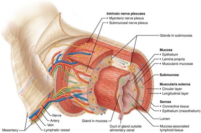
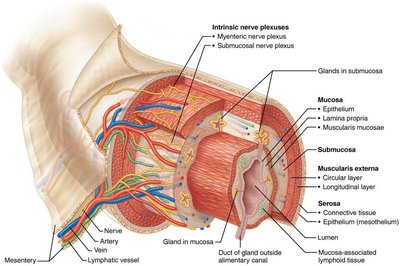
All digestive organs share four basic layers:
Mucosa: Lines the lumen; functions in secretion, absorption, and protection. Composed of epithelium, lamina propria, and muscularis mucosae.
Submucosa: Areolar connective tissue containing blood vessels, lymphatics, and nerve plexuses.
Muscularis Externa: Responsible for segmentation and peristalsis; consists of inner circular and outer longitudinal muscle layers.
Serosa: Outermost layer; visceral peritoneum. In the esophagus, replaced by fibrous adventitia.
Blood Supply and Control of the Digestive System
Splanchnic Circulation
Splanchnic Circulation: Includes arteries branching from the aorta (hepatic, splenic, gastric, mesenteric) and hepatic portal circulation, which delivers nutrient-rich blood from digestive organs to the liver for processing.
Enteric Nervous System
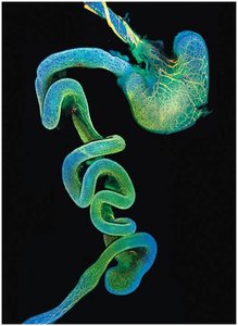
The GI tract has its own nervous system, the enteric nervous system (gut brain), which contains more neurons than the spinal cord. It controls motility and consists of:
Submucosal Nerve Plexus: Regulates glands and smooth muscle in the mucosa.
Myenteric Nerve Plexus: Controls GI tract motility.
Neural Reflex Pathways
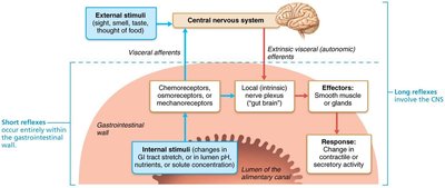
Short Reflexes: Mediated by enteric nerve plexuses; respond to stimuli within the GI tract.
Long Reflexes: Respond to stimuli inside or outside the gut, involving the autonomic nervous system. Parasympathetic enhances digestion; sympathetic inhibits it.
Regulation of Digestive Activity
Mechanical and Chemical Stimuli: Receptors in GI tract walls respond to stretch, osmolarity, pH, and presence of substrates.
Effectors: Smooth muscle and glands mix and move contents, and secrete digestive juices or hormones.
Control: Intrinsic (enteric nervous system) and extrinsic (autonomic nervous system) neurons, as well as hormones, regulate digestive activity.
Functional Anatomy of the Digestive System
Mouth and Associated Organs
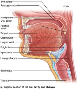
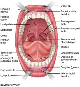
The mouth (oral cavity) is the entry point for food, where it is chewed and mixed with saliva. Associated organs include the tongue, salivary glands, and teeth.
Boundaries: Lips, cheeks, palate, and tongue.
Epithelium: Stratified squamous, with keratinized areas for protection.
Tongue
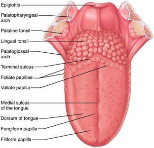
The tongue is composed of skeletal muscle and functions in gripping, repositioning, mixing food, forming the bolus, and initiating swallowing, speech, and taste.
Intrinsic Muscles: Change shape of the tongue.
Extrinsic Muscles: Alter tongue position.
Papillae: Filiform (friction, no taste buds), fungiform (taste buds), vallate (taste buds), foliate (taste buds).
Terminal Sulcus: Divides oral cavity portion (body) from oropharynx portion (root).
Salivary Glands
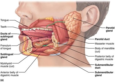
Salivary glands produce saliva, which cleanses the mouth, dissolves food chemicals, moistens food, and begins starch breakdown with amylase.
Major Glands: Parotid, submandibular, and sublingual.
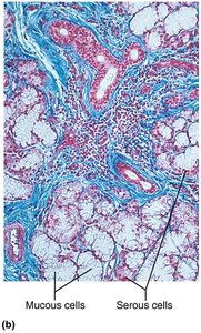
Secretory Cells: Serous (watery, enzymes) and mucous (mucus).
Composition: Mostly water, slightly acidic, contains electrolytes, enzymes, proteins, and antimicrobial agents.
Control: Parasympathetic stimulation increases salivation; sympathetic decreases it.
Teeth
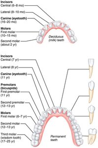
Teeth are essential for mastication (chewing), breaking food into smaller fragments. Dentition includes primary (deciduous) and permanent teeth, classified as incisors, canines, premolars, and molars.
Dental Formula: Indicates number and position of teeth.
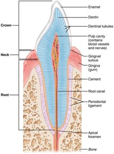
Structure: Crown (enamel-covered), neck, root (embedded in jawbone), cement, periodontal ligament, gingival sulcus, dentin, pulp cavity, root canal, apical foramen.
Tooth and Gum Disease
Dental Caries: Demineralization of enamel and dentin due to bacterial action; prevention includes flossing and brushing.
Gingivitis: Inflammation of gums due to plaque and tartar; reversible with treatment.
Periodontitis: Advanced gum disease; immune response damages tissues and bone, increasing risk for heart disease and stroke.
Clinical Notes
Ankyloglossia: Short lingual frenulum causing "tongue-tied" condition; treated surgically.
Xerostomia: Dry mouth due to insufficient saliva; can result from medications or diseases.
Impacted Tooth: Tooth trapped in jawbone, often wisdom teeth; treated by surgical removal.
Primary Teeth Care: Essential for oral health and proper development of permanent teeth.
Layer | Main Function | Key Features |
|---|---|---|
Mucosa | Secretion, absorption, protection | Epithelium, lamina propria, muscularis mucosae |
Submucosa | Support, vascular supply | Areolar connective tissue, blood vessels, lymphatics, nerve plexus |
Muscularis externa | Motility (peristalsis, segmentation) | Inner circular, outer longitudinal muscle layers |
Serosa | Protection, structural support | Visceral peritoneum, mesothelium |
Type of Tooth | Function | |
|---|---|---|
Incisor | Cutting | |
Canine | Tearing/Piercing | |
Premolar | Grinding/Crushing | |
Molar | Grinding |
